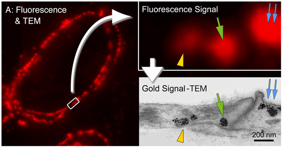
Learn more about our FluoroNanogold™ products...
By combining gold and fluorescence into one immunoprobe, the same specimen may be imaged using both fluorescence microscopy (e.g., with a confocal microscope) and at the ultrastructural level by electron microscopy.
|
|||||||||||||||||||||||||||||||||||||||||||||||||||||||||||||||||||||||||||||||||||||||||||||
Alexa Fluor®* 647 FluoroNanogold™ 2-in-1 immunolabeling for Super-Res! |
|
 |
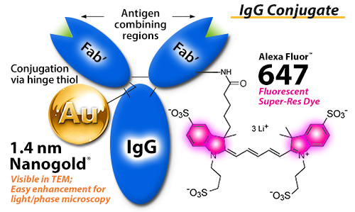
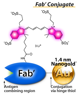
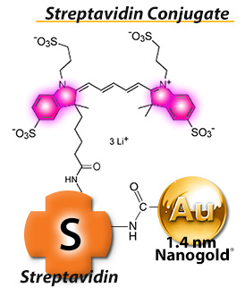
Simultaneously label for Super-Res Each secondary antibody / Fab' includes TWO labels
Finally-- Put your Super-Res images into cellular context!
|
 |
 |
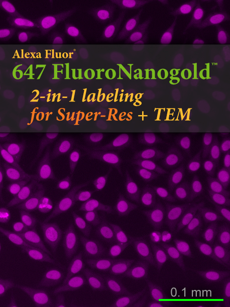
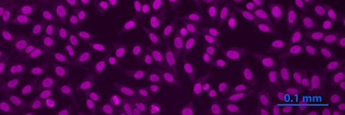 Fluorescent labeling of Alexa Fluor® 647 and Nanogold® - Fab' tertiary probe. The specimen is a slide from the NOVA Lite ANA HEp-2 test, an indirect immunofluorescent test system for the screening and semi-quantitative determination of anti-nuclear antibodies (ANA) in human serum (see ). The slide was stained using positive pattern control human sera, a Mouse anti-Human secondary antiboidy, and combined Alexa Fluor® 647 and Nanogold® - Fab' tertiary probe. Specimens were washed with PBS (30 minutes) between each step, then blocked by the addition of 7 % nonfat dried milk to the tertiary antibody solution (original magnification 400 X). |
 |
Alexa Fluor® 647 FluoroNanogold™ |
Alexa Fluor® 647 FluoroNanogold™ |
 Alexa Fluor®* 488 FluoroNanogold™
Our new Alexa Fluor® FluoroNanogold™ conjugates will give you greatly enhanced performance compared with conventional fluorophores. Benefit from greatly improved fluorescence properties, combined with a new level of freedom from background and non-specific binding.
- Increased fluorescence signal and higher quantum yield - ideal for scarce targets or dynamic systems where exposure needs to be restricted.
- Fluorescence remains high and consistent across wider pH range.
- Improved solubility means reduced non-specific interactions, lower background and higher signal-to-noise ratios.
- Uses fluorescein filter sets.
- Available in 1 mL or affordable 0.5 mL sizes.
![[Alexa Fluor® 488 FluoroNanogold: structure and fluorescence staining] (48k)]](../Images/catfig15.jpg)
Left: Structure of Alexa Fluor® 488 and Nanogold® - Fab', showing covalent attachment of components.
Right: Fluorescent staining obtained using combined combined Alexa Fluor® 488 and Nanogold® - Fab' tertiary probe. The specimen is a slide from the NOVA Lite ANA HEp-2 test, an indirect immunofluorescent test system for the screening and semi-quantitative determination of anti-nuclear antibodies (ANA) in human serum (see ). The slide was stained using positive pattern control human sera, a Mouse anti-Human secondary antiboidy, and combined Alexa Fluor® 488 and Nanogold® - Fab' tertiary probe. Specimens were washed with PBS (30 minutes) between each step, then blocked by the addition of 7 % nonfat dried milk to the tertiary antibody solution (original magnification 400 X).
Alexa Fluor® 488 FluoroNanogold™-Fab' |
Alexa Fluor® 488 FluoroNanogold™-Streptavidin |

 Alexa Fluor®* 546 FluoroNanogold™
Our new Alexa Fluor® FluoroNanogold™ conjugates will give you greatly enhanced performance compared with conventional fluorophores. Benefit from greatly improved fluorescence properties, combined with a new level of freedom from background and non-specific binding.
- Increased fluorescence signal and higher quantum yield - ideal for scarce targets or dynamic systems where exposure needs to be restricted.
- Fluorescence remains high and consistent across wider pH range.
- Improved solubility means reduced non-specific interactions, lower background and higher signal-to-noise ratios.
- Uses fluorescein filter sets.
- Available in 1 mL or affordable 0.5 mL sizes.
Alexa Fluor® 546 FluoroNanogold™-Fab'
|
Alexa Fluor® 546 FluoroNanogold™-Streptavidin
|
 Alexa Fluor®* 594 FluoroNanogold™
Our Alexa Fluor®* 594 FluoroNanogold™conjugates offer an a second fluorophore, enabling the labeling of more than one target within a specimen with the brightness and photostability of the Alexa Fluor® reagents.
- Use for multiple labeling: distinguish a FluoroNanogold™-labeled target from a second target labeled with fluorescein, Alexa Fluor® 488, green fluorescent protein, or other fluorophores.
- Uses Texas Red filter sets.
- Available in 1 mL or affordable 0.5 mL sizes.
![[Alexa Fluor® 594 Labeling (64k)]](../Images/catfig17.jpg)
Localization of caveolin-1a in ultrathin cryosection of human placenta using a new FNG; caveolin 1 alpha is primarily located to caveolae in placental endothelial cells. One-to-one correspondence is found between fluorescent spots and caveola labeled with gold particles (right). Ultrathin cryosections collected on formvar film-coated nickel EM grids were incubated with chicken anti-human caveolin-1a IgY for 30 min at 37°C, then with biotinylated goat anti-chicken F(ab)2 (13 mg/ml) for 30 min at 37°C, then stained with ALEXA-594 FluoroNanogold™-Streptavidin (1:50 dilution) for 30 min at room temperature. Non-specific sites on cryosections were blocked with 1% milk - 5% fetal bovine serum-PBS for 30 minutes at room temperature (figure courtesy of T. Takizawa, Ohio State University, Columbus, OH).
![[Alexa Fluor® 594 Conjugates (46k)]](../Images/catfig16.gif)
Structure of Alexa Fluor®* 594 FluoroNanogold™Fab' and Streptavidin conjugates, showing covalent attachment.
Alexa Fluor® 594 FluoroNanogold™-Fab' |
Alexa Fluor® 594 FluoroNanogold™-Streptavidin |
 Fluorescein FluoroNanogold™
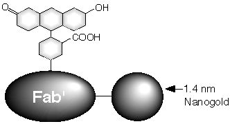
Fluorescein is the most widely used fluorophore; fluorescein FluoroNanogold™conjugates combine fluorescein and Nanogold® in a single convenient, stable probe.
|
 |
FluoroNanogold™Staining of Microtubules
![[FluoroNanogold labeling of Microtubules by fluorescent, light and electron microscopy] (302k)](../Images/catfig6.jpg) |
| Immunolocalization of microtubules with Fluorescein FluoroNanogold™-Fab' in the same human monocyte visualized by various microscopies: (A) Fluorescence; (B) Phase; (C) DIC, and (D) Electron Microscopy (with silver enhancement). (Micrographs courtesy of Dr. J. M. Robinson and Dr. D. Vandré, Ohio State University). |
Fluorescein FluoroNanogold™-Fab' |
Fluorescein FluoroNanogold™-Streptavidin |
Custom Labeling
We can also create custom conjugates, using your own primary antibody (IgG or Fab'), proteins, peptides, lectins, and other molecules. Please use our Custom Synthesis Form to let us know your needs, or drop us a line by phone or email at tech@nanoprobes.com.
* Alexa Fluor is a registered trademark of Life Technologies, Inc.
| © 1990-2017 Nanoprobes, Inc. All rights reserved. | Sitemap |


