 |
Nanoprobes, Incorporated
95 Horseblock Road, Unit 1, Yaphank, NY 11980-9710
Tel: (877) 447-6266 (Toll-Free in US) or (631) 205-9490 | Fax: (631) 205-9493
tech@nanoprobes.com | www.nanoprobes.com |
Updated: February, 2011
NANOGOLD® PRODUCT INFORMATION
Alexa Fluor®* 546 FluoroNanoGold™**
- Streptavidin
[Alexa Fluor®* 546-FluoroNanogoldTM**-Streptavidin]

Product
Number |
Product Name |
| 7416 |
Alexa Fluor® 546 FluoroNanogold™-Streptavidin |
| Appearance: |
Pale pink liquid |
| Revision: |
1.1 (February 2011) |
Technical Assistance Online
![[2025-PDF]](../Images/pdf.gif) Instructions (PDF) Instructions (PDF)
Material Safety Data Sheet (PDF) --coming soon
Congratulations on your acquisition of a dual labeling cytochemical reagent: Alexa Fluor® FluoroNanogold™-Streptavidin. This unique streptavidin probe contains both the 1.4 nm Nanogold® particle and Alexa Fluor®, both covalently bound, enabling both fluorescence and electron microscope observation of the exact same structure in a single labeling procedure. This probe is smaller than a whole IgG molecule, does not aggregate, and fluorescence quenching due to the gold particle is low. Alexa Fluor® has been found to have significant advantages over fluorescein or rhodamine: it is brighter and quenches less readily.
CONTENTS
Warning: For research use only. Not recommended or intended for diagnosis of disease in humans or animals. Do not use internally or externally in humans or animals. Non radioactive and non carcinogenic.
PRODUCT INFORMATION
Alexa Fluor® 546 FluoroNanogold™ is a unique, dual-purpose probe developed specially for confocal and electron microscopy.1
Alexa Fluor®546 FluoroNanogold™-Streptavidin consists of streptavidin conjugated to both Alexa Fluor® 546 dye and the 1.4 nm Nanogold® particle.2 A streptavidin molecule labeled with Alexa Fluor® 546 and Nanogold® is shown in Figure 1.
In the fluorescence microscope, these probes may be used just like conventional fluorescently-labeled streptavidin,3 while in the electron microscope they are visualized in exactly the same manner as for Nanogold® conjugates.4,5 The combination of the fluorescent and electron microscopy, referred as correlative microscopy,16 allows imaging of the same exact structure in both microscopes.
The covalent label linkage is stable indefinitely.
These reagents are supplied at a concentration of 0.08 mg/mL of streptavidin dissolved in 20 mM phosphate buffered saline (150 mM NaCl) at pH 7.4 (PBS), with 0.1% BSA and 0.05% sodium azide as preservatives.
FluoroNanogold™ conjugates should be stored at 2-8°C. DO NOT FREEZE. PROTECT FROM LIGHT.
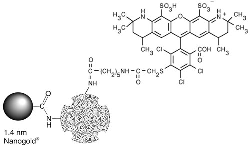 |
| Figure 1: Streptavidin covalently conjugated to Alexa Fluor® 546 and Nanogold® to give Alexa Fluor® Fluoro-Nanogold™ - Streptavidin. |
PROPERTIES
- Alexa Fluor® FluoroNanogold™ contains an extremely uniform 1.4 nm diameter gold particle (±10%) and 2 – 3 fluorophores per streptavidin.
- The absorption maximum of Alexa Fluor® 546 occurs near 556 nm, and the emission maximum is near 573 nm. These values are very similar to those of the Cy3 dye.
- The Alexa Fluor® 546 dye is readily excited by the 546 nm line of the mercury-arc lamp and the 543 nm line of the He-Ne laser.
- Alexa Fluor® FluoroNanogold™- Streptavidin is smaller than a single whole IgG molecule. It has a similar size as Fab'- Nanogold®, the smallest gold immunoprobe commercially available, and will penetrate and reach antigens inaccessible to other gold probes.
- Alexa Fluor® FluoroNanogold™- Streptavidin is chromatographically purified through gel filtration columns. There are absolutely no aggregates or other molecular weight impurities. This is in sharp contrast to colloidal gold conjugates which usually are prepared by centrifugation to remove the largest aggregates, and frequently contain smaller aggregates.
- Close to 1 Nanogold® label to 1 Streptavidin molecule make this product distinct from the 0.2 - 10 variable stoichiometry of other colloidal gold preparations.
- The Alexa Fluor® FluoroNanogold™ streptavidin is used as a secondary antibody. It may also be used as a tertiary labeled antibody, or a custom Alexa Fluor® FluoroNanogold™ conjugate may be the primary antibody. If additional antibody incubation steps are used, rinse with buffer 3 (3 X 10 mins) after incubation.
- Alexa Fluor® FluoroNanogold™- Streptavidin particles do not have affinity to proteins as do colloidal golds. This reduces background and false labeling. Alexa Fluor® FluoroNanogold™- Streptavidin develops better with silver than do most colloidal golds giving it higher sensitivity.
- Both silver and gold enhancement can be used to make the labeling useful for electron microscopy and light microscopy.
GENERAL CONSIDERATIONS FOR IMMUNOSTAINING WITH FLUORONANOGOLD™ REAGENTS
Basically, normal methodologies for each component of the label may be used successfully with Alexa Fluor® FluoroNanogold™ labeling agents.
Due to some quenching of fluorescence by the gold particle, slightly higher concentrations of streptavidin are recommended for incubations.
A blocking agent of 5% non-fat dried milk has been found to reduce nonspecific background staining in some cases: this should be used before incubation with probe (in standard wash/blocking steps), and additionally, the Alexa Fluor® FluoroNanogold™- Streptavidin probe can be diluted in a solution also containing 1% non-fat dried milk before it is applied.
USING EM STAINS WITH ALEXA FLUOR® FLUORONANOGOLD™
Because the 1.4 nm Alexa Fluor® FluoroNanogold™ particles are so small, over staining with OsO4, uranyl acetate or lead citrate may tend to obscure direct visualization of individual Nanogold® particles.
Four recommendations for improved visibility of Alexa Fluor® FluoroNanogold™ are:
- Use of reduced amounts or concentrations of usual stains.
- Use of lower atomic number stains such as NanoVan™, a Vanadium based stain.6
- Enhancement of Alexa Fluor® FluoroNanogold™ with silver developers, such as LI Silver™ or HQ Silver™.
- Enhancement of Alexa Fluor® FluoroNanogold™ with the gold developer, GoldEnhance™.
THIOL CAUTION
Nanogold® particles experience loss of gold clusters (Nanogold®) upon exposure to thiols such as ß-mercaptoethanol (BME) or dithiothreitol (DTT).
Avoid use of thiol agents.
If a reducing environment is needed, reduce the protein, then purify from the thiol agent by column chromatography. Use non-metallic columns, and include 5 mM EDTA with the eluent, since trace metals catalyze thiol oxidation back to disulfides; most thiols do not reoxidize within several hours to several days following this procedure. Then use the Alexa Fluor® FluoroNanogold™.
If a reducing agent is absolutely required, use a non-thiol agent, such as TCEP (triscarboxyethyl phosphine).
TEMPERATURE CAUTION
Although Nanogold® is stable under most conditions,7 labeled specimens or conjugates may not be stable above 80°C for long periods. Best results are obtained at room temperature or 4°C.
It is best to use silver or gold enhancement before procedures requiring temperatures above 37°C, such as baking, or use low temperature embedding media (e.g., Lowicryl) if labeling before embedding.8
METHODS and PROTOCOLS
Several publications describe the successful application of FluoroNanogold™ for light and electron microscopy. These provide additional protocols, details and applications that may be helpful in obtaining the best results (Refs. 1, 15-19).
IN SITU HYBRIDIZATION WITH ALEXA FLUOR® FLUORONANOGOLD®-STREPTAVIDIN
PROTOCOL
(adapted from that of Hacker, G.W., et al.).9,10
Practical considerations:
This is a robust and reliable technique for routine use. It is intended for biotinylated hybridization probes. Other types of reporter molecules may be demonstrated by application of a biotinylated linking antibody system. The sensitivity of Nanogold-silver ISH depends, to a large degree, at the dilution of FluoroNanogold™-Streptavidin and the duration of silver development applied. Careful adjustment of the protease predigestion is necessary. In some preparations, some degree of unwanted background staining in connective tissue is obtained. This is in part due to fixation and possible excessive protease treatment and can often be reduced by application of higher dilutions of Alexa Fluor-FluoroNanogold™-Streptavidin.
Fluorescence microscopy should be performed before silver enhancement since the silver particles quench fluorescence.
- Deparaffinize sections from formaldehyde-fixed tissue in fresh xylene (2 times 15 min each).
- Rinse and rehydrate in graded alcohols and distilled water (2-3 min each).
- Soak in phosphate-buffered saline (PBS, 20mM, pH 7.6) for 3 min.
- Incubate with 0.1 mg/mL proteinase K (code no. 1 373 196, from Boehringer Mannheim, Mannheim, FRG) in PBS at 37°C for about 8 min. The duration is critical and has to be tested very carefully, depending on tissue, fixation and other factors.
- Rinse in 2 changes of PBS, 3 min.
- Permeabilize with 0.3% Triton X-100 in PBS for 15 min.
- Wash in PBS for 2 min.
- Rinse in 2 changes of distilled water, dehydrate with graded alcohols (50%, 70%, 98% isopropanol) for 1 min each and air-dry the sections.
- Prehybridize with 1:1 mixture of deionized formamide and 20% dextran sulfate in 2X SSC at 50 °C for 5 min.
- Carefully shake off excess prehybridization block.
- Add one drop of biotinylated DNA probe on the section and cover with a small coverslip. Avoid air bubbles.
- Heat sections on heating block at 92-94 °C for 8-10 min to denature DNA.
- Incubate in a moist chamber at 37°C overnight (or for at least 2 hours).
- Post-hybridization washes (5 min each): 2 changes of 4X SSC (1st wash to remove coverslips), 2X SSC, 0.1X SSC, 0.05X SSC, and then distilled water.
- Put slides into Lugol's iodine solution (Merck, Darmstadt, Germany) for 5 min.
- Wash in tap water and then distilled water.
- Put into 2.5% sodium thiosulfate for a few seconds until sections are colorless. Then wash in tap water for 5 min and distilled water for 2 min.
- Immerse in PBS containing 0.1% fish gelatin (45% concentrate – Cat. No. G-7765, Sigma-Aldrich, Steinheim, Germany) and 0.1% Tween-20 for 5 min.
- Incubate sections with FluoroNanogold™-Streptavidin diluted 1:200 to 1:500 in PBS containing 1% BSA at room temperature for 60 min.
- Wash in 3 changes of PBS containing 0.1% fish gelatin and 0.1% Tween-20 for 5 min each.
- Repeatedly wash in distilled water for at least 10 min altogether, the last 2 rinses in ultrapure water (EM-grade).
- The specimen may now be observed by fluorescence microscopy.
- Perform silver or gold Enhancement (e.g., LI Silver™, HQ Silver™ or GoldEnhance™), as specified in those product directions.
- Rinse carefully in tap water for at least 3 min. After silver amplification, sections can be counterstained with Nuclear Fast Red, dehydrated and mounted in Permount or in DPX (BDH Chemicals, Poole, UK). Do not use Eukitt.
Solutions:
- Phosphate-buffered saline (PBS): 10X PBS (Mg2+ and Ca2+-free) pH 7.6: 11.36 g Na2HPO4, 2.72 g KH2PO4, 87.0 g NaCl in 800 mL distilled water. Adjust pH with concentrated NaOH and add distilled water to a final volume of 1 L.
- Standard Sodium Citrate Buffer (SSC): 175.32 g NaCl and 88.23 g sodium citrate in 800 ml distilled water. Adjust pH with NaOH to 7.0 and add distilled water to a final volume of 1 L.
FLUORESCENCE MICROSCOPY IMMUNOLABELING
WITH ALEXA FLUOR® FLUORONANOGOLD™-STREPTAVIDIN
PROTOCOL
If aldehyde-containing reagents have been used for fixation, these should be quenched before labeling. This may be achieved by incubating the specimens for 5 minutes in 50 mM glycine solution in PBS (pH 7.4). Ammonium chloride (50 mM) or sodium borohydride (0.5 - 1 mg/ml) in PBS may be used instead of glycine.
The procedure below3 describes an example of the use of a Alexa Fluor® FluoroNanogold™ -Streptavidin as a secondary antibody probe.
Dilutions of Alexa Fluor®-FluoroNanogold™-Streptavidin will vary with different procedures, but a final concentration of 0.2 – 10 μg/mL is advisable as a starting point for most applications; for simultaneous electron microscopy labeling, a compromise between the optimum concentrations for fluorescence and electron microscopy maybe necessary.
Other protocols and techniques used with fluorescently-labeled antibodies or streptavidin may also be used with Alexa Fluor® FluoroNanogold™ conjugates.
- Fix cells in freshly-prepared 2% formaldehyde in PBS for 15 mins at 20°C; alternatively, fix in 100% methanol at -20°C for 3 minutes; if methanol fixation is used, skip to step 4.
- Wash in PBS (3 x 10 mins).
- Permeabilize in 0.2% Triton X-100 plus 1% normal serum (NS) in PBS at pH 7.4 for 5 minutes on ice.
- Wash in PBS with 1% NS (3 X 10 mins).
- Incubate in the appropriate concentration of biotinylated primary antibody for 1 hour at room temperature in a humidified chamber.
- If using 22 mm X 22 mm square cover slips, 30 μL of diluted antibody is placed on the coverslip and the coverslip is inverted onto a glass slide. The slide is then placed in a humidified chamber which is incubated at room temperature.
- Alternatively, a tertiary labeling procedure may be used where the primary antibody is not biotinylated, but the second antibody is.
- If additional antibody incubation steps are used, rinse with buffer 3 (3 X 10 mins) after incubation.
- Wash in PBS with 1% NS + 5% non-fat dried milk (3 X 10 mins).
- Incubate with Alexa Fluor®-FluoroNanogold™-Streptavidin reagent at a concentration of 0.2 – 10 μg/mL (diluted in buffer containing 1% non-fat dried milk) for 1 hour in a humidified chamber at room temperature.
- Wash in PBS (4 X 10 mins).
- Mount coverslip with a drop of mounting medium. Observe as usual.
PBS Buffer:
20 mM phosphate
150 mM NaCl
pH 7.4
ELECTRON MICROSCOPY WITH ALEXA FLUOR® FLUORONANOGOLD™- STREPTAVIDIN
The procedures given in this section are complete immunolabeling procedures, and are also recommended for Nanogold® conjugates.
If the specimen has already been labeled and observed by fluorescence microscopy, it requires only mounting, silver or gold enhancement (usually necessary) and staining according to your usual electron microscopy protocol before observation.
If aldehyde-containing reagents have been used for fixation, these should be quenched before labeling. This may be achieved by incubating the specimens for 5 minutes in 50 mM glycine solution in PBS (pH 7.4). Ammonium chloride (50 mM) or sodium borohydride (0.5 - 1 mg/ml) in PBS may be used instead of glycine.
Cells in Suspension
If the cells are already labeled, mount, stain and observe as usual. If a different specimen is to be used, the procedure below is recommended:
PROTOCOL FOR CELLS IN SUSPENSION
- Optional fixing of cells: e.g., with glutaraldehyde (0.05 - 1% for 15 minutes) in PBS. Do not use Tris buffer since this contains an amine which reacts with glutaraldehyde.
- Centrifuge cells (e.g. 1 ml at 107 cells/mL) at 300 X g, 5 minutes; discard supernatant; resuspend in 1 mL buffer. Repeat this washing (centrifugation and resuspension) 2 times.
- Incubate cells with 0.02 M glycine in PBS (5 mins). Centrifuge, then resuspend cells in PBS-milk buffer (specified below) or PBS containing 1 % BSA for 5 minutes.
- Place 50 - 200 μL of cells into Eppendorf tube and add 5 - 10 μL of biotinylated primary antibody. Incubate 30 minutes with occasional shaking (do not create bubbles which will denature proteins). Alternatively, a tertiary labeling procedure may be used where the primary antibody is not biotinylated, but the second antibody is. If additional antibody incubation steps are used, rinse with PBS-milk buffer (3 X 10 mins) after incubation.
- Wash cells using PBS-Milk as described in step 2 (2 X 5 mins). Resuspend in 1 mL PBS-Milk buffer.
- Dilute Alexa Fluor®-FluoroNanogold™-Streptavidin to a concentration of 0.2 – 10 μg/mL in PBS-Milk buffer and add 30 μL to cells; incubate for 30 minutes with occasional shaking.
- Wash cells in PBS buffer as described in step 2 (2 X 5 mins).
- Fix cells and antibodies using a final concentration of 1% glutaraldehyde in PBS for 15 minutes. Then remove fixative by washing with PBS buffer (3 X 5 mins).Centrifuge cells (e.g. 1 ml at 107 cells/mL) at 300 X g, 5 minutes; discard supernatant; resuspend in 1 mL buffer. Repeat this washing (centrifugation and resuspension) 2 times.
PBS-Milk Buffer:
20 mM phosphate
150 mM NaCl
pH 7.4
1% Non-fat dried milk (final concentration)
PBS Buffer:
20 mM phosphate
150 mM NaCl
pH 7.420 mM phosphate pH 7.4
Optional, may reduce background:
0.5 M NaCl
0.05% Tween 20
0.1% gelatin (high purity)
Negative Staining
Negative staining may be used for electron microscopy of small structures or single molecules which are not embedded. Negative stain must be applied after the silver enhancement.
NanoVan™ negative stain is specially formulated for use with Nanogold® reagents;6 it is based on vanadium, which gives a lighter stain than uranium, lead or tungsten-based negative stains and allows easier visualization of Alexa Fluor® FluoroNanogold™ particles with little or no silver enhancement.
Other procedures may be used; for example the FluoroNanogold™ reagent may be used as a tertiary labeled probe in a system where a biotinylated secondary antibody is used with an unlabeled primary antibody. If additional antibody incubation steps are used, rinse with PBS-Milk (3 X 5 mins) after incubation.
Thin Sections
Labeling with Alexa Fluor® FluoroNanogold™-Streptavidin may be performed before (the pre-embedding method)9,10 or after embedding and sectioning (the post-embedding method).9,10 The procedures for both methods are described below.
Thin sections mounted on grids are floated on drops of solutions on parafilm or in well plates. Hydrophobic resins usually require pre-etching.
PROTOCOL FOR PRE-EMBEDDING METHOD:9
If specimen has already been labeled with Alexa Fluor® FluoroNanogold™-Streptavidin, skip to step 9. If a fresh specimen is required for EM, the following procedure is recommended.
- Float on a drop of water for 5 - 10 minutes.
- Incubate cells with 1% bovine serum albumin in PBS buffer at pH 7.4 for 5 minutes; this blocks non-specific protein binding sites and minimizes non-specific antibody binding.
- Incubate with biotinylated primary antibody, diluted at usual working concentration in PBS-milk buffer or PBS containing 1% BSA (1 hour or usual time. Buffer formulations are given below). Alternatively, a tertiary labeling procedure may be used where the primary antibody is not biotinylated, but the second antibody is. If additional antibody incubation steps are used, rinse with buffer 3 (3 X 10 mins) after incubation.
- Rinse with PBS-Milk (3 X 1 min).
- Incubate with Alexa Fluor®-FluoroNanogold™-Streptavidin reagent diluted to a concentration of 0.2 – 10 μg/mL in PBS- Milk with 1% normal serum for 10 minutes to 1 hour at room temperature.
- Rinse with PBS- Milk (3 X 1 min), then PBS (3 X 1 min).
- Postfix with 1% glutaraldehyde in PBS (10 mins).
- Rinse in deionized water (2 X 5 min).
- Perform silver or gold enhancement (e.g., HQ Silver™ or GoldEnhance™), as specified in those product directions.
- Dehydrate and embed according to usual procedure.
- Stain (uranyl acetate, lead citrate or other staining reagent) as usual before examination.
PROTOCOL FOR POST-EMBEDDING METHOD:9
- Prepare sections on plastic or carbon-coated nickel grid. Float on a drop of water for 5 - 10 minutes.
- Incubate with 1% solution of bovine serum albumin in PBS buffer at pH 7.4 for 5 minutes to block non-specific protein binding sites.
- Incubate with biotinylated primary antibody, diluted at usual working concentration in PBS-Milk buffer or PBS containing 1% BSA (1 hour or usual time. Buffer formulations are given below). Alternatively, a tertiary labeling procedure may be used where the primary antibody is not biotinylated, but the second antibody is. If additional antibody incubation steps are used, rinse with buffer 3 (3 X 10 mins) after incubation.
- Rinse with PBS-Milk (3 X 1 min).
- Incubate with Alexa Fluor® FluoroNanogold™-Streptavidin reagent diluted to a concentration of 0.2 – 10 μg/mL in PBS- Milk with 1% normal serum for 10 minutes to 1 hour at room temperature.
- Rinse with PBS (3 X 1 min).
- Postfix with 1% glutaraldehyde in PBS at room temperature (3 mins).
- Rinse in deionized water for (2 X 5 min).
- If desired, contrast sections with uranyl acetate and/or lead citrate before examination.
Silver or gold enhancement may also be used to render the Alexa Fluor® FluoroNanogold™ particles more easily visible (see below); this is recommended if stains such as uranyl acetate or lead citrate are applied. Silver or gold enhancement should be completed before these stains are applied.
PBS-Milk Buffer:
20 mM phosphate
150 mM NaCl
pH 7.4
1% Non-fat dried milk (final concentration)
PBS Buffer:
20 mM phosphate
150 mM NaCl
pH 7.420 mM phosphate pH 7.4
Optional, may reduce background:
0.5 M NaCl
0.05% Tween 20
0.1% gelatin (high purity)
SPECIAL CONSIDERATIONS FOR DIRECT VIEWING OF ALEXA FLUOR® FLUORONANOGOLD™ IN THE ELECTRON MICROSCOPE
For most work, silver or gold enhancement is recommended to give a good signal in the electron microscope (see below). For particular applications, such as high resolution cryo-electron microscopy, visualization of the Alexa Fluor® FluoroNanogold™ directly may be desirable. Generally this requires very thin samples and precludes the use of other stains.
Alexa Fluor® FluoroNanogold™-Streptavidin provides a much improved resolution and smaller probe size over other colloidal gold conjugate products. However, because Nanogold® is only 1.4 nm in diameter, it will not only be smaller, but will appear less intense than, for example, a 5 nm gold particle. With careful work, however, Nanogold® may be seen directly through the binoculars of a standard EM even in 80 nm thin sections. However, achieving the high resolution necessary for this work may require new demands on your equipment and technique.
Several suggestions follow:
- Before you start a project with Alexa Fluor® FluoroNanogold™-Streptavidin it is helpful to see it so you know what to look for. Dilute the Alexa Fluor® FluoroNanogold™ stock 1:5 and apply 4 µL to a grid for 1 minute. Wick the drop and wash with deionized water 4 times.
- View Alexa Fluor® FluoroNanogold™-Streptavidin at 100,000 X magnification with 10 X binoculars for a final magnification of 1,000,000 X. Turn the emission up full and adjust the condenser for maximum illumination.
- The alignment of the microscope should be in order to give 0.3 nm resolution. Although the scope should be well aligned, you may be able to skip this step if you do step 4.
- Objective stigmators must be optimally set at 100,000 X. Even if the rest of the microscope optics are not perfectly aligned, adjustment of the objective stigmators may compensate and give the required resolution. You may want to follow your local protocol for this alignment but since it is important, a brief protocol is given here:
- At 100,000 X (1 X 106 with binoculars), over focus, under focus, then set the objective lens to in focus. This is where there is the least amount of detail seen.
- Adjust each objective stigmator to give the least amount of detail in the image.
- Repeat steps a and b until the in focus image contains virtually no contrast, no wormy details, and gives a flat featureless image.
- Now underfocus slightly, move to a fresh area, and you should see small black dots of 1.4 nm size. This is the Nanogold®. For the 1:5 dilution suggested, there should be about 5 to 10 gold spots on the small viewing screen used with the binoculars. Contrast and visibility of the gold clusters is best at 0.2 - 0.5 microns defocus, and is much worse at typical defocus values of 1.5 - 2.0 microns commonly used for protein molecular imaging.
- In order to operate at high magnification with high beam current, thin carbon film over fenestrated holey film is recommended. Alternatively, thin carbon or 0.2% Formvar over a 1000 mesh grid is acceptable. Many plastic supports are unstable under these conditions of high magnification/high beam current and carbon is therefore preferred. Contrast is best using thinner films and thinner sections.
- Once you have seen Nanogold® you may now be able to reduce the beam current and obtain better images on film. For direct viewing with the binoculars reduction in magnification from 100,000 X to 50,000 X makes the Nanogold® much more difficult to observe and not all of the gold particles are discernable. At 30,000 X (300,000 X with 10 X binoculars) Nanogold® particles are not visible. It is recommended to view at 100,000 X, with maximum beam current, align the objective stigmators, and then move to a fresh area, reduce the beam, and record on film.
- If the demands of high resolution are too taxing or your sample has an interfering stain, or is thick, a very good result may be obtained using silver or gold enhancement to give particles easily seen at lower magnification.
SILVER ENHANCEMENT OF ALEXA FLUOR® FLUORONANOGOLD™ FOR EM
Alexa Fluor® FluoroNanogold™ will nucleate silver deposition resulting in a dense particle 2-80 nm in size or larger depending on development time. It should be completed before any staining reagents such as osmium tetroxide, lead citrate or uranyl acetate are applied, since these will nucleate silver deposition in the same manner as gold and produce non-specific staining. Silver development is recommended for applications of Alexa Fluor® FluoroNanogold™-Streptavidin in which stains are to be used, otherwise the Alexa Fluor® FluoroNanogold™-Streptavidin particles may be difficult to visualize against the stain.
Our LI Silver™ silver enhancement system is convenient and not light sensitive, and suitable for all applications. Improved results in the EM may be obtained using HQ Silver™, which is formulated to give slower, more controllable particle growth and more uniform particle size distribution.13
Specimens must be thoroughly rinsed with deionized water before silver enhancement reagents are applied. This is because the buffers used for antibody incubations and washes contain chloride ions and other anions which form insoluble precipitates with silver. These are often light-sensitive and will give non-specific staining. To prepare the developer, warm the components to room temperature and mix equal amounts of the components immediately before use. Nanogold™ will nucleate silver deposition resulting in a dense particle 2-20 nm in size or larger depending on development time. Use of nickel grids is sometimes preferred.
Fluorescence microscopy should be performed BEFORE silver enhancement. This is because the silver-enhanced gold particles can quench fluorescence.
The relevant procedure for immunolabeling should be followed. Silver enhancement is then performed as follows:
SILVER ENHANCEMENT PROTOCOL
- Rinse with deionized water (2 X 5 mins).
- OPTIONAL (may reduce background):5 Wash several times with 0.02 M sodium citrate buffer, pH 7.0.
- Float grid with specimen on freshly mixed developer for 1-12 minutes, or as directed in the instructions for the silver reagent. More or less time can be used to control particle size. A series of different development times should be tried, to find the optimum time for your experiment. With HQ Silver™, a development time of 6 min. usually gives 15-40 nm round particles.
- Rinse with deionized water (3 X 1 min).
- Mount and stain as usual.
Fixing with osmium tetroxide may cause some loss of silver; if this is found to be a problem, slightly longer development times may be appropriate. Alternatively, use of 0.1% osmium tetroxide instead of 1% has been found to give similar levels of staining while greatly reducing etching of the silver particles.
NOTE: Treatment with osmium tetroxide followed by uranyl acetate staining can lead to loss of the silver enhanced Nanogold® particles. This may be prevented by gold toning:14
GOLD TONING PROTOCOL
- After silver enhancement, wash thoroughly with deionized water.
- 0.05% gold chloride: 10 minutes at 4°C.
- Wash with deionized water.
- 0.5% oxalic acid: 2 mins at room temperature.
- 1% sodium thiosulfate (freshly made) for 1 hour.
- Wash thoroughly with deionized water and embed according to usual procedure.
GOLD ENHANCEMENT OF ALEXA FLUOR® FLUORONANOGOLD™-STREPTAVIDIN FOR EM
The small 1.4 nm Nanogold® particles may alternatively be enhanced (grown to a larger size) for better visibility using GoldEnhance™ EM (catalog number 2113), which catalytically deposits gold around the Nanogold®, making a larger solid gold particle. Gold enhancement may be preferable to silver enhancement in some cases due to the different properties of GoldEnhance™ EM:
GOLD ENHANCEMENT PROTOCOL
- Gold is chemically more stable and is not depleted by osmium or uranyl stains;
- Gold has higher backscattering and is useful for SEM;
- GoldEnhance™ EM is not light insensitive – it can be used in normal room lighting, and development followed in the light microscope;
- GoldEnhance™ EM may be used with physiological buffers, such as ones containing chloride, which precipitates silver enhancers.
GoldEnhance™ EM follows a similar procedure to silver enhancement. For specific directions, see those that accompany GoldEnhance™ EM.
REFERENCES
- Hainfeld, J. F.; Furuya, F. R.; Powell, R. D., and Liu, W.: Combined ALEXA-488 and Nanogold Antibody Probes. Microsc. Microanal., 8, (Suppl. 2: Proceedings) (Proceedings of Microscopy and Microanalysis 2002); Voekl, E.; Piston, D.; Gauvin, R.; Lockley, A. J.; Bailey, G. W., and McKernan, S., Eds.; Cambridge University Press, New York, NY, p. 1030CD (2002).
- Hainfeld, J. F., and Powell R. D.: Nanogold Technology: New Frontiers in Gold Labeling. Cell Vision, 4, 308-324 (1997); Furuya, F. R., and Hainfeld, J. F.: A 1.4nm Gold cluster covalently attached to antibodies improves immunolabeling. J. Histochem. Cytochem., 40, 177-184 (1992); Furuya, F. R.; Hainfeld, J. F., and Powell, R. D.: A new 1.4 nm Gold-Fab' Probe. Proc. 49th Ann. Mtg., Electron. Micros. Soc. Amer.; Bailey, G. W. (Ed.), San Francisco Press, San Francisco, CA, p. 284 (1991).
- Spector, D. L., and Smith, H. C.: Redistribution of U-snRNPs during mitosis. Exp. Cell. Res., 163, 87-94 (1986).
- Powell, R. D.; Hainfeld, J. F.; Halsey, C. M. R.; Joshi, V. N.; Hacker, G. W.; Hauser-Kronberger, C., and Takvorian, P. M.: Combined Cy3 / Nanogold Conjugates for Immunocytochemistry and In Situ Hybridization. Microsc. Microanal., 5, (Suppl. 2: Proceedings) (Proceedings of Microscopy and Microanalysis 1999); G. W. Bailey, W. G. Jerome, S. McKernan, J. F. Mansfield, and R. L. Price (Eds.); Springer-Verlag, New York, NY, 478-479 (1999).
- Powell, R. D.; Halsey, C. M. R., and Hainfeld, J. F.: Combined fluorescent and gold immunoprobes: Reagents and methods for correlative light and electron microscopy. Micros. Res. Technique, 42, 2-12 (1998); Powell, R. D.; Halsey, C. M. R.; Spector, D. L.; Kaurin, S. L.; McCann, J., and Hainfeld, J. F.: A covalent fluorescent-gold immunoprobe: "simultaneous" detection of a pre-mRNA splicing factor by light and electron microscopy. J. Histochem. Cytochem., 45, 947-956 (1997); Robinson, J. M., and Vandré, D. D.: Efficient immunocytochemical labeling of leukocyte microtubules with FluoroNanogold: An important tool for correlative microscopy. J. Histochem. Cytochem. 45, 631-642 (1997).
- Tracz, E.; Dickson, D. W.; Hainfeld, J. F., and Ksiezak-Reding, H.: Paired helical filaments in corticobasal degeneration: the fine fibrillary structure with NanoVan. Brain Res., 773, 33-44 (1997); Gregori, L.; Hainfeld, J. F.; Simon, M. N., and Goldgaber, D.: Binding of amyloid beta protein to the 20S proteasome. J. Biol. Chem., 272, 58-62 (1997); Hainfeld, J. F.; Safer, D.; Wall, J. S.; Simon, M. N.; Lin, B. J., and Powell, R. D.: Methylamine Vanadate (NanoVan) Negative Stain. Proc. 52nd Ann. Mtg., Micros. Soc. Amer.; G. W. Bailey and Garratt-Reed, A. J., (Eds.); San Francisco Press, San Francisco, CA, p. 132 (1994).
- Hainfeld, J. F., and Furuya, F. R.: Silver-enhancement of Nanogold and undecagold. in Immunogold-Silver Staining: Principles, Methods and Applications (M. A. Hayat, Ed.), CRC Press, Boca raton, FL., 1995: pp. 71-96.
- Krenács, T., and Krenács, L.: Comparison of embedding media for immunogold-silver staining. in Immunogold-Silver Staining: Principles, Methods and Applications (M. A. Hayat, Ed.), CRC Press, Boca raton, FL., 1995: pp. 57-69.
- Hacker, G. W., Hauser-Kronberger, C.; Zehbe, I.; Su, H.; Schiechl, A.; Dietze, O., and Tubbs, R.: In Situ localization of DNA and RNA sequences: Super-sensitive In Situ hybridization using Streptavidin-Nanogold®-Silver Staining: Minireview, Protocols and Possible Applications. Cell Vision, 4, 54-65 (1997).
- Zehbe, I.; Hacker, G. W.; Su, H.; Hauser-Kronberger, C.; Hainfeld, J. F., and Tubbs, R.: Sensitive in situ hybridization with catalyzed reporter deposition, streptavidin-Nanogold, and silver acetate autometallography. Detection of single-copy human papillomavirus. Am. J. Pathol., 150, 1553-1561 (1997).
- J. E. Beesley, in Colloidal Gold: Principles, Methods and Applications (M. A. Hayat, Ed.), Academic Press, New York, 1989; Vol. 1, pp. 421-425.
- Lujan, R.; Nusser, Z.; Roberts, J. D. B.; Shigemoto R.; Ohishi, H., and Somogyi, P.: Differential plasma membrane distribution of metabotropic glutamate receptors mGluR1 alpha, mGluR2 and mGluR5, relative to neurotransmitter release sites. J. Chem. Neuroanat., 13, 219-241 (1997).
- Humbel, B. M.; Sibon, O. C. M.; Stierhof, Y.-D., and Schwarz, H.: Ultra-small gold particles and silver enhancement as a detection system in immunolabeling and In Situ hybridization experiments; J. Histochem. Cytochem., 43, 735-737 (1995).
- Arai, R.; Geffard, M.; and Calas, A. Intensification of labeling of the immunogold silver staining method by gold toning. Brain Res. Bull., 28, 343-345 (1992).
- Robinson, J. M.; Takizawa, T.; Pombo, A., and Cook, P. R.: Correlative fluorescence and electron microscopy on ultrathin cryosections: bridging the resolution gap. J. Histochem. Cytochem., 49, 803-808 (2001).
- Robinson, J. M.; Takizawa, T., and Vandre, D. D.: Applications of gold cluster compounds in immunocytochemistry and correlative microscopy: comparison with colloidal gold. J. Microsc., 199, 163-179 (2000).
- Takizawa, T., and Robinson, J. M.: FluoroNanogold is a bifunctional immunoprobe for correlative fluorescence and electron microscopy. J. Histochem. Cytochem., 48, 481-486 (2000).
- Takizawa, T., and Robinson, J. M.: Analysis of antiphotobleaching reagents for use with FluoroNanogold in correlative microscopy. J. Histochem. Cytochem., 48, 433-436 (2000).
- Takizawa, T., Suzuki, K., and Robinson, J. M.: Correlative microscopy using FluoroNanogold on ultrathin cryosections. Proof of principle. J. Histochem. Cytochem., 46, 1097-1102 (1998).
* Alexa Fluor is a registered trademark of Molecular Probes, Inc.
** Patented technology.
Questions? Technical Support is available. Drop us a line-- we're here to help.
|
Nanoprobes, Incorporated
95 Horseblock Road, Unit 1, Yaphank, NY 11980-9710
Tel: (877) 447-6266 (Toll-Free in US) or (631) 205-9490 | Fax: (631) 205-9493
tech@nanoprobes.com | www.nanoprobes.com |
|