|
Advantages over silver:
|

Our customers report superb results in their labs, |
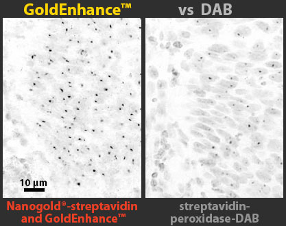
Much better than enzymatic detection for LM!
|
Direct detection: (Micrographs courtesy of Prof. G. W. Hacker. Medical Research Coordination Center, University of Salzburg, Austria) |
See our paper on Gold-Based Autometallography. |
 GoldEnhance™ EM Plus - Improved formula
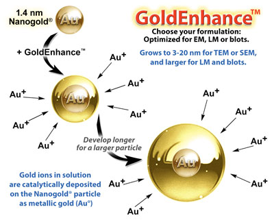
Deposit metallic gold to enlarge ultrasmall gold nanoparticles for EM - Use it with Nanogold®!
- Slow enhancement for an easy control of particle size
- Great for SEM: Gold gives a much better backscatter signal than silver
- Low background - No autonucleation for 40 minutes
- Neutral pH for best ultrastructural preservation
- Permanent staining: does not fade
- Resistant to osmium tetroxide etching: may safely be used before osmium tetroxide staining with no need for gold toning or other protective treatments. Gold is stable in the oxidizing agent OsO4; silver would dissolve.
- Compatible with physiological buffers and other halide solutions --these would precipitate silver.
- Can be used for specimens on metal surfaces (e.g. cell culture substrates)
- Low viscosity
- Light insensitive
- Mix-and-use
- Our new favorite for EM!
Protocol: Pre-embedding Nanogold® labeling
with enhancement by GoldEnhance™ EM PlusInstructions and technical information
Instructions (PDF)
Material Safety Data Sheet (PDF)
 GoldEnhance™ EM (original)
Our original gold enhancer for EM.
- High density of enlarged particles for clear imaging.
- Resistant to osmium tetroxide etching: may safely be used before osmium tetroxide staining with no need for gold toning or other protective treatments (silver is dissolved by the oxidizing agent OsO4; gold is stable).
- for SEM, gold gives a much better backscatter signal than silver.
Instructions and technical information
Instructions (PDF)
Material Safety Data Sheet (PDF)
 GoldEnhance™ LM
Formulated for robust and clear development for light microscopy.
- Autonucleation is minimal even after 1-2 hours - more convenient for multiple samples, or if sample access is restricted (e.g. automated processing).
- Much faster than chemiluminescence.
- Observe with brightfield optics - simpler and less expensive than fluorescence.
- Permanent staining: does not fade.
Instructions and technical information
Instructions (PDF)
Material Safety Data Sheet (PDF)
 GoldEnhance™ Blots
Specially formulated for protein blots. Each kit will stain 12 (7 x 8.4 cm) Westerns.
- Fast development. Observe 10 pg antigen within 10 minutes.
- Higher sensitivity than silver. Able to detect 5 pg in immuno-dot blots.
- Generates a dark purple, permanent stain. No fading!
- No precipitation with physiological buffers or halides.
- Consistent, reproducible results.
Instructions and technical information
Instructions (PDF)
Material Safety Data Sheet (PDF)
|
| © 1990-2017 Nanoprobes, Inc. All rights reserved. | Sitemap |


