|
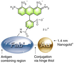
Nanoprobes collaborator Dr. Peter Takvorian has pioneered the use of FluoroNanogold™ to study microsporida, a class of spore-forming protozoans, responsible for many tropical diseases as well as some of the opportunistic infections associated with the human immunodeficiency virus (HIV).
 Microsporidial infection is usually diagnosed by the identification and morphology or proteins that make up the polar tube, the apparatus by which these organisms inject their infective sporoplasm into host cells. Microsporidial infection is usually diagnosed by the identification and morphology or proteins that make up the polar tube, the apparatus by which these organisms inject their infective sporoplasm into host cells.
Complete diagnosis can be a complex procedure requiring both light and electron microscopy, this presenting a natural opportunity for FluoroNanogold™.
Recently, Dr. Takvorian's consortium partners, Dr. Ann Cali of Rutgers and Dr. Louis Weiss of Albert Einstein College of Medicine, continued this work on the fluorescent and electron microscopic localization of novel structural components of the polar tube and spore wall in Encephalitozoon cuniculi microsporida identified using a shotgun proteomic strategy. In doing so, they made a new discovery: one such protein, the 51-kDa ECU10_1500, also forms a filamentous network in the lumen of the parasitophorous vacuole.6 A similar network was also observed in another species, Encephalitozoon hellem. It is not yet clear what role this network plays in the infective process, but as these components are discovered, and their roles established, they present new drug targets for these difficult-to-treat pathogens.
E. hellem was chosen for analysis by immunoelectron microscopy because its parasitophorous vacuoles were more frequently stained using the anti-ECU10_1500 antibody than were those of E. cuniculi. Infected RK13 host cell cultures grown on poly-D-lysine plus laminin-coated glass coverslips were fixed with 2% formaldehyde in Dulbecco’s modified phosphate-buffered saline (DPBS) for 30 minutes at room temperature, then permeabilized for 20 minutes in 0.2% Triton X-100 in DPBS, and blocked overnight in 3% BSA / 0.2% Triton X-100 in DPBS at 4°C. Immunostaining was performed in 1% BSA / 0.2% Triton X-100 in DPBS. Fixed host cell cultures were incubated with 1 : 1,000 dilutions of mouse antisera to recombinant parasite proteins at 37°C for 1.5 hours, then with a 1:50 dilution of Alexa Fluor 488 FluoroNanogold™ for 25 minutes at 37°C and 35 minutes at room temperature. Coverslips were mounted with antifade mounting medium and cured overnight in the dark before fluorescence observation.
For electron microscopic observation, cultures were postfixed in 2% formaldehyde in DPBS for 3 minutes at room temperature and washed three times in DPBS and three times in water. FluoroNanogold™-stained cultures were then enhanced using LI Silver for 15 or 25 minutes then checked by brightfield optical microscopy. Cultures were dehydrated in graded ethanols and embedded in situ in Epon resin. Glass coverslips were removed by treatment with hydrofluoric acid.7 Thin sections were cut with a microtome, mounted on Formvar-coated grids, stained with uranyl acetate, and observed in the transmission electron microscope.
This finding may lead to new drugs to combat opportunistic infections associated with HIV as well as other hard-to-treat tropical diseases.
The combination of correlative microscopy with proteomics provides a powerful platform to extend this approach to other systems.
- Takizawa, T.; Suzuki, K., and Robinson, J. M.: Correlative Microscopy Using FluoroNanogold on Ultrathin Cryosections: Proof of Principle; J. Histochem. Cytochem., 46, 1097-1102 (1998).
- Takizawa, T., and Robinson, J. M.: Correlative Microscopy of Ultrathin Cryosections is a Powerful Tool for Placental Research. Placenta, 24, 557-565 (2003).
- Schröder-Reiter, E.; Houben, A.; Grau, J, and Wanner, G.: Characterization of a peg-like terminal NOR structure with light microscopy and high-resolution scanning electron microscopy. Chromosoma, 115, 50-59. (2006).
- McRae, R.; Lai, B.; Vogt, S., and Fahrni, C. J.: Correlative microXRF and optical immunofluorescence microscopy of adherent cells labeled with ultrasmall gold particles. J. Struct. Biol., 155, 22-29 (2006).
- Iliev, A. I.; Ganesan, S.; Bunt, G., and Wouters, F. S.: Removal of pattern-breaking sequences in microtubule binding repeats produces instantaneous tau aggregation and toxicity. J. Biol. Chem., 281, 37195-37204 (2006).
- Ghosh, K.; Nieves, E.; Keeling, P.; Pombert, J. F.; Henrich, P. P.; Cali, A., and Weiss, L. M.: Branching network of proteinaceous filaments within the parasitophorous vacuole of Encephalitozoon cuniculi and Encephalitozoon hellem. Infect Immun., 79, 1374-1385 (2011).
- Abstract from the American society for Microbiology
- Powell, R. D.; Halsey, C. M. R.; Spector, D. L.; Kaurin, S. L.; McCann, J., and Hainfeld, J. F. A covalent fluorescent-gold immunoprobe: "simultaneous" detection of a pre-mRNA splicing factor by light and electron microscopy. J. Histochem. Cytochem., 45, 947-956 (1997).
|
Also in this issue:
|