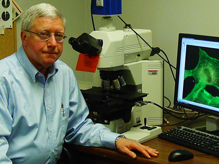
Dr. John M. Robinson of Ohio State University and collaborator Dr. Toshihiro Takizawa are two of the pre-eminent FluoroNanogold™ users, providing the first proof of principal of truly correlative microscopy.
John’s specialty is the ultrastructure of the placenta; like the membranes discussed in our last issue, the placental plasma membrane is a delicate organelle, and in order to study it at the molecular level, he has specialized in the development of methods for Nanogold® and FluoroNanogold™ labeling in ultrathin cryosections. Among the targets successfully localized are the caveolins such as that shown in our main article.
Want to hear more? Dr. Robinson will give a talk on Correlative Microscopy, including FluoroNanogold™, at the Histochemistry Society's Histochemistry 2012, to be held from March 21 to 24, 2012, in Woods Hole, MA.

Dr. John Robinson examines a cell labeled with a fluorescent cytoskeletal probe.
| “ |
Correlative microscopy using tissue samples presents problems not found with single cell preparations. |
|
| ...FluoroNanogold™ was key to our success... |
| |
...in high-resolution immunofluorescence and correlative immunoelectron microscopy of tissues using ultrathin cryosections as the immunolabeling substratum. |
" |
--Dr. John Robinson
Ohio State University, Dept. of Physiology and Cell Biology