 |
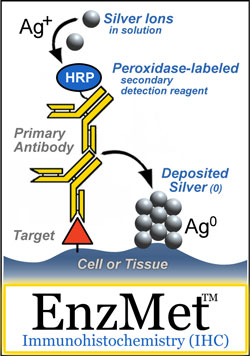
In addition to being the best staining and detection method for in situ hybridization, EnzMet™ is also one of the best staining and detection methods for immunohistochemistry (IHC).
EnzMet™ combines high sensitivity with precision, leaving the cellular morphology more visible and allowing easy differentiation from other stains.
Best of all, these results are obtained with a simple new substrate - like DAB, but better.
EnzMet™ can yield better results with a simple monomeric immunoperoxidase than using organic chromogens such as DAB with premium polymerized immunoperoxidase reagents, metal enhancement reagents, target or signal amplification methods. |
EnzMet provides several important
features and advantages for immunostaining:
- Extraordinary selectivity combined with virtually zero background provides highest possible contrast.
Your biomarkers will stand out with super clarity.
- Virtually no diffusion of reaction product means super-sharp signals.
Target distribution is defined at the highest possible resolution. Low abundance markers are visualized by punctuate staining for accurate localization and quantitation.
- Black, sharply defined, non-diffusing stain lets you clearly see underlying morphology.
EnzMet is readily distinguished from most counterstains. Double staining is easy: a second target stained with a conventional organic chromogen is readily differentiated.
- EnzMet works by deposition of silver, so it also provides high contrast for electron microscopy (EM).
Samples can be processed for EM without further immunostaining. Transfer your specimens to EM for ultrastructural localization, or use EnzMet for correlative brightfield light and electron microscopy.

Comparison of EnzMet with conventional DAB immunohistochemistry and enhanced DAB, showing superior contrast and sensitivity even without further amplification. Cytokeratins in paraffin-embedded bladder tumor stained with monoclonal primary antibody (AE1/AE3, Dako) and peroxidase secondary with (A) EnzMet;(B) DAB; (C) DAB enhanced using nickel (II); and (D) Polymerized HRP-secondary (Envision, Dako) developed with DAB.
|
Outside Evaluation of EnzMet™: The Findings
Evaluation of EnzMet for immunohistochemical application was conducted by Tubbs, Pettay and co-workers, using high-complexity tissue microarrays (TMAs).[1]
88 common solid tumors were evaluated by enzyme metallography (EnzMet) using an automated slide staining system (Ventana Medical Systems); targets were chosen to assess the ability of EnzMet to specifically localize encoded antigens in the nucleus (estrogen receptor), cytoplasm (cytokeratins), and cytoplasmic membrane (HER2) in TMAs.
The intensity of staining for all three antigens evaluated was comparable for breast tumors as well as carcinomas of kidney, colon, and prostate. However, the quality of staining in EnzMet IHC preparations was much sharper, and the stain deposits were better defined, showing a more punctate appearance, than that found with DAB.
Full concordance was found between the EnzMet and conventional IHC results. The EnzMet reaction product was dense and sharply defined, did not appreciably diffuse, and provided excellent high-resolution differentiation of cellular compartments in paraffin sections for the nuclear, cytoplasmic, and cell membrane-localized antigens evaluated.
The higher density of elemental silver deposited during enzyme metallography permitted evaluation of core immunophenotypes at a relatively low magnification, without the need for oil immersion, allowing more tissue to be screened in an efficient manner.
In addition, the signal is stable and provided a permanent record.
Concordance data are shown below:
| Tumor Type |
Enzyme Metallography (EnzMet™) |
Conventional DAB IHC |
| |
HER2 |
Cytokeratin (CK) |
Estrogen Receptor (ER) |
HER2 |
Cytokeratin (CK) |
Estrogen Receptor (ER) |
| Breast carcinoma, invasive ductal |
3/15 |
15/15 |
9/15 |
3/15 |
15/15 |
9/15 |
| Breast carcinoma, invasive lobular |
0/15 |
15/15 |
11/15 |
0/15 |
15/15 |
11/15 |
| Prostate, adenocarcinoma |
0/15 |
12/15 |
0/15* |
0/15 |
12/15 |
0/15* |
| Colon, adenocarcinoma |
0/15 |
15/15 |
0/15* |
0/15 |
15/15 |
0/15* |
| Renal cell carcinoma |
0/15 |
0/15 |
0/15* |
0/15 |
0/15 |
0/15* |
Data are given as cores positive/total cores.
*Epithelial tumor cells negative; occasional stromal cells positive by both DAB and EnzMet IHC.
We believe that EnzMet will have just as much impact on IHC for clinical pathology as it has on ISH.
EnzMet™ also remains a remarkably versatile and sensitive reagent for light microscopy in general, and for this application, you can still purchase EnzMet™ directly from Nanoprobes.
- Tubbs R.; Pettay J.; Powell R.; Hicks D. G.; Roche P.; Powell W.; Grogan T., and Hainfeld, J. F.: High-resolution immunophenotyping of subcellular compartments in tissue microarrays by enzyme metallography. Appl. Immunohistochem. Mol. Morphol., 13, 371-375 (2005).
- Abstract from the Applied Immunohistochemistry and Molecular Morphology
- Powell, R. D.; Hainfeld, J. F.; Gutierrez, E.; Furuya, F. R.; Joshi, V. N.; Liu, W., and Takvorian, P. M.: Nanogold Labels, Covalent Gold, and Enzyme Metallography as Components of Nanodevices. AIP Conf. Proc. (Fritzsche, W., and Bier, F., Eds.), 1062, 91-99 (2008).
- Abstract from the AIP Conference Proceedings.
|
Also in this issue:
|