Our mission
Nanoprobes is a small biotech company dedicated to developing nanoparticles for research and cancer therapy.
Cancer is an unrelenting foe.
We have tried many approaches but despite some promising preliminary results, until now none have overcome it. Cancer is a complex adversary, and patients with no effective recourse have continued to die.
Read on to learn about the decades of research that brought us to our current breakthrough in our quest to cure cancer.

In memoriam
Dr. Wenqiu Liu, PhD
Our former Production Manager and Core Team Member
This work is dedicated to Dr. Wenqiu Liu, who died of triple negative breast cancer at the age of 57.
At Nanoprobes, all profits go toward finding a cancer cure.
Donate to "Project Cure Cancer"
A short history of our research
–in the words of Dr. Jim Hainfeld, our founder and chief research scientist, who has devoted his life and career of over 50 years to cancer research.
“We believe that the nanoparticle approach, which is our specialty, may be able to contribute to some new and better cancer diagnostics or treatments. A few of our contributions are listed below. We hope this research has stimulated other scientists to ultimately benefit patients.”
Much of this work was done in collaboration with our great friend and colleague, Dr. Henry Smilowitz, at the University of Connecticut Health Center.
Gold nanoparticles enhance radiotherapy
More than 50% of cancer victims undergo radiotherapy (RT). While radiation kills tumor cells more than normal cells (they’re more sensitive when dividing), normal cells are in the beam path and take a hit. This limits the radiation that can be safely given, so even though it shrinks tumors, it is usually not curative.
What if we could increase the absorption of the radiation in the tumor to deposit more energy there?
Gold is a good radiation absorber, so we tried to deliver gold nanoparticles (AuNPs) to tumors to boost its effectiveness there. We tried this in mice and indeed, it led to many of them being cured (Fig.1).
Read the article we published on our findings: “The use of gold nanoparticles to enhance radiotherapy in mice” at https://doi.org/10.1088/0031-9155/49/18/n03. This article has been cited by other research papers 2207 times.
A cure to cancer? As mentioned, cancer is a formidable foe. Although high atomic number (Z) nanoparticles are even being tried clinically to enhance RT, we found some serious drawbacks to this approach. High Z elements best absorb at kilovolt X-ray energies, whereas most cancer RT is done at million volt energy X-rays for better penetration to deep tumors. The high Z particles then absorb ~1,000 times less and the enhancement is largely lost. 2. At the high dose needed for best results the nanoparticles are taken up by macrophages in liver, spleen, and skin and because the particles are dark, white mice turned black for even a year.
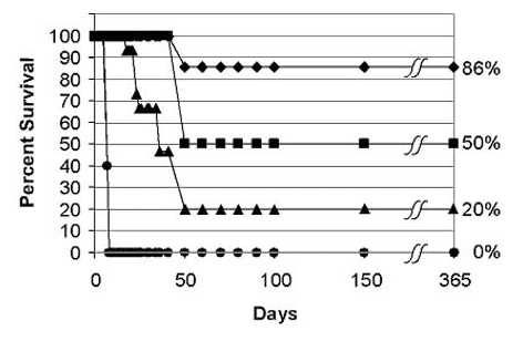
Fig. 1. After intravenous gold nanoparticle injection, the gold loaded tumors and made the radiotherapy more effective. 0% survived 1 year with no treatment, 20% with RT alone, 50% with AuNPs, and 86% with a double dose of AuNPs.
High atomic number (Z) nanoparticles like gold can be used to better image cancer
Because of their high X-ray absorption, high Z nanoparticles if targeted to tumors could image them better for earlier detection or for RT treatment planning (where to direct the X-rays). We first showed this in mice revealing their blood vessels and cancer (Fig.2).
Read the article: “Gold nanoparticles: a new X-ray contrast agent” https://doi.org/10.1259/bjr/13169882 (Cited 1813 times by other research papers).
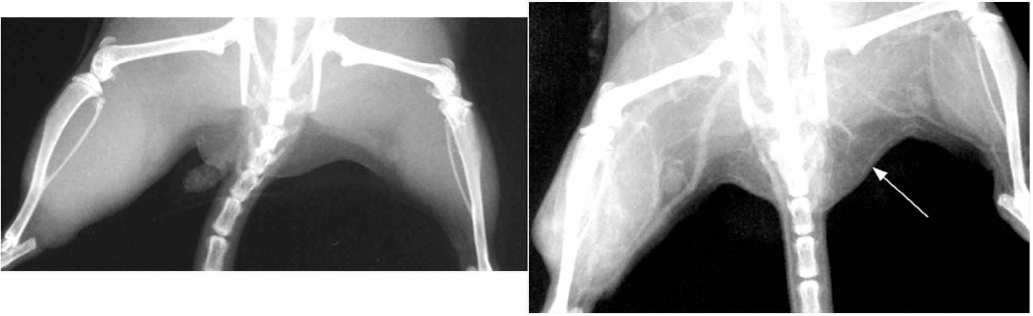
Fig. 2. Left: X-ray of mouse legs. Right: 2 minutes after intravenous injection of gold nanoparticles making small vessels visible (arrow) and increased vascularity seen in the brighter leg on the left where a tumor is growing.
Curing brain cancer in mice with Gold NPs + RT
Brain tumors could be imaged at high resolution (Fig. 3) and combined with RT produced 50% long-term survival (Fig. 4).
Read the article: “Gold nanoparticle imaging and radiotherapy of brain tumors in mice” https://doi.org/10.2217/nnm.12.165
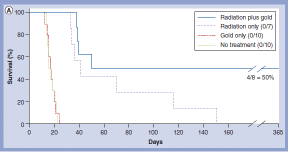
Fig. 4. RT+AuNPs cured 50% of mice with brain tumors.
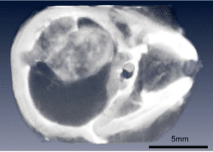
Fig. 3. Brain tumor in mouse clearly imaged after IV injection of gold nanoparticles.
Gold nanoparticles precisely visualize, quantify tumors on Micro CT
High atomic number (Z) nanoparticles like gold can be used to better image cancer.
The microlocalization of antibody-targeted gold nanoparticles could be assessed using calibrated microCT imaging of live mice (Fig. 5).
Read the article: Micro-CT enables microlocalisation and quantification of Her2-targeted gold nanoparticles within tumour regions. https://doi.org/10.1259/bjr/42612922
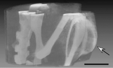
Figure 5. Micro-CT volume images from live mouse with Her2+ tumor (arrow). Imaging was 20 h after iv injection of Herceptin–15 nm AuNPs. Bar 55 mm.
IV-injected gold NPs load brain tumors better than via direct injection
Intravenously-injected gold nanoparticles accessed intracerebral F98 rat gliomas better than AuNPs infused directly into the tumor site by convection enhanced delivery.
Which is better: direct intratumoral injection or intravenous injection? We answered this question by this study. IV application was superior (Fig. 6).
Read the article: “Intravenously-injected gold nanoparticles (AuNPs) access intracerebral F98 rat gliomas better than AuNPs infused directly into the tumor site by convection enhanced delivery”. https://doi.org/10.2147/ijn.s154555
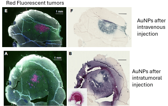
Fig. 6. Intravenous AuNPs better target brain tumors than injecting them directly into the tumors.
Antibody Targeted Gold Nanoparticles heated with Near Infrared Ablated Tumors
By controlling the aggregation of spherical AuNPs, their absorption could be shifted to the infrared (IR) and thus heated with an infrared laser. We showed that anti-EGFr antibody targeted AuNPs irradiated with an IR laser could “cure” all tumors (Fig. 7).
Read the article: “Infrared-Transparent Gold Nanoparticles Converted by Tumors to Infrared Absorbers Cure Tumors in Mice by Photothermal Therapy” https://doi.org/10.1371/journal.pone.0088414
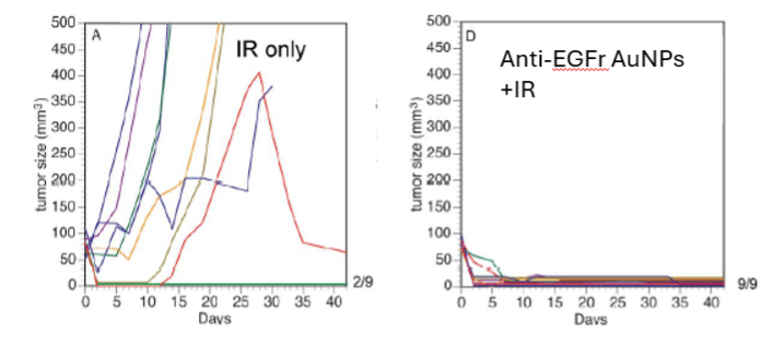
Fig. 7. Infrared heating alone ablated 2 of 9 tumors (left graph), but if given after IV anti-EGF AuNPs that aggregated in the tumor, 100% of tumors (9/9) were eradicated.
Infrared Heated Gold Nanoparticles Enhance Radiotherapy
Infrared transparent 15 nm gold nanoparticles were intratumorally injected and then became infrared absorptive due to their aggregation by the low pH of the tumor (Fig. 8). The tumor was then heated with a halogen lamp filtered to peak at 820n to 48°C for 5 min, then immediately irradiated with X-rays. For an equivalent effect the X-ray dose could be reduced 3.7 times if combined with pre heating.
Read the article: “Gold nanoparticle hyperthermia reduces radiotherapy dose” https://doi.org/10.1016/j.nano.2014.05.006
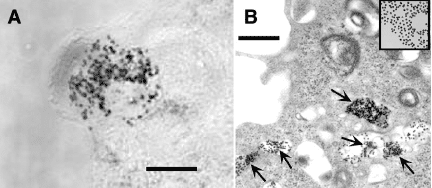
Figure 8. (A) Light micrograph of the in vitro uptake and aggregation of lipoic acid AuNPs in the cytoplasm of one tumor cell. Bar = 10 μm.
Iodine Nanoparticles improved treatment of brain cancers in mice
To counter the negative effect of long-term skin discoloration we developed colorless iodine nanoparticles which also could better detect tumors (Fig. 9).
Treatment with X-rays greatly extended life (Fig. 10).
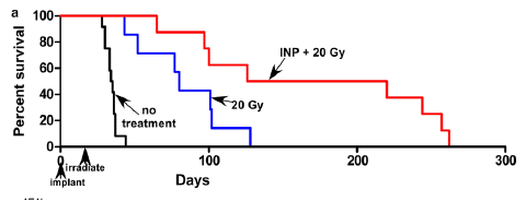
Fig. 10. Survival graph showing improved survival of human glioma (in mouse brain) with RT+Iodine Nanoparticles.(INPs).
Read the articles: “Iodine nanoparticle radiotherapy of human breast cancer growing in the brains of athymic mice” https://doi.org/10.1038/s41598-020-72268-0
and “Iodine nanoparticles enhance radiotherapy of intracerebral human glioma in mice and increase efficacy of chemotherapy”. https://doi.org/10.1038/s41598-019-41174-5
However, once again these results were with low energy X-rays and would not work as well with standard 6MV (MegaVoltage) or 10MV X-rays.
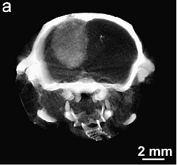
Fig. 9. MicroCT image of brain tumors after IV injection of iodine nanoparticles.
Radiotherapy of brain tumor resection cavity edge enhanced with Xoft balloon irradiator: A Monte Carlo study.
Removal of brain tumors often leaves tumor cells at the periphery. By inserting a balloon with a 50kVp needle X-ray source (Xoft, Inc) this periphery can be irradiated. Gold or iodine nanoparticles would boost the effectiveness.
Read the article: “Accelerated brachytherapy with the Xoft electronic source used in association with iodine, gold, bismuth, gadolinium, and hafnium nano-radioenhancers” https://doi.org/10.1016/j.brachy.2022.06.008
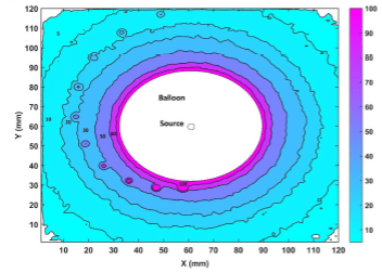
Fig. 11. MC calculated isodose curves representing absorbed dose distribution.
Barium Titanate Nanoparticles Enhance MV X-rays
In continuing the search for nanoparticles that enhance MV X-rays we developed biocompatible barium titanate nanoparticles. These accumulated in tumors after IV injection, and did not discolor the skin.
In collaboration with Dr. Smilowitz at the University of Connecticut Health Center these NPs were shown to enhance MV X-rays in mice with tumors. Presumably the titanate component generates free radicals when impinged with X-rays.
Magnetic Nanoparticles Ablate Tumors by heating in an alternating magnetic field (AMF).
We developed biocompatible magnetic nanoparticles that selectively loaded tumors. When exposed to an AMF they heat up, enough to damage the tumor (Figs. 12, 13).
We found tumors could be heated to 50°C in as little as 5 sec. Although some mice could be “cured”, the equipment for treating humans is not available and the high level injected discolored skin for months and had poor clearance.
Read about it: “Intravenous Magnetic Nanoparticle Hyperthermia” https://doi.org/10.2147/ijn.s43770
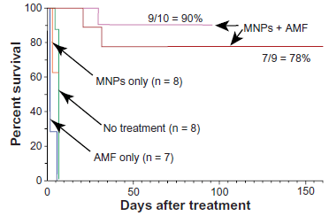
Fig. 13. Survival greatly improved after IV magnetic nanoparticles (MNPs) and AMF.
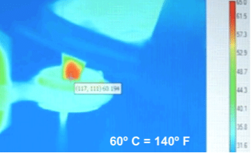
Fig. 12. Thermal camera image of tumor on leg of mouse being heated when immersed in an alternating magnetic field.
Developed a Breast Cancer Test now being used worldwide
In our quest to make nanoparticles useful tools for more sensitive detection we discovered a way for the enzyme horseradish peroxidase to deposit silver, producing a bold signal that can be easily seen by microscopy. We called this Enzyme Metallograph, or EnzMet for short.
A good test for Her2 breast cancer was needed by pathologists to identify this type of cancer from a biopsy. We worked with the Cleveland Clinic and applied our EnzMet to see the Her2 gene duplication in a biopsy, thus identifying that cancer (Fig. 14).
This test was shown to Roche and the results are so distinctive and definitive that they licensed the technology from Nanoprobes. Subsequently the test was approved by the FDA and is being used worldwide. Based on this test, patients that are Her2 positive will go on a monoclonal antibody therapy called Herceptin.
Learn more: Saving Lives with EnzMet
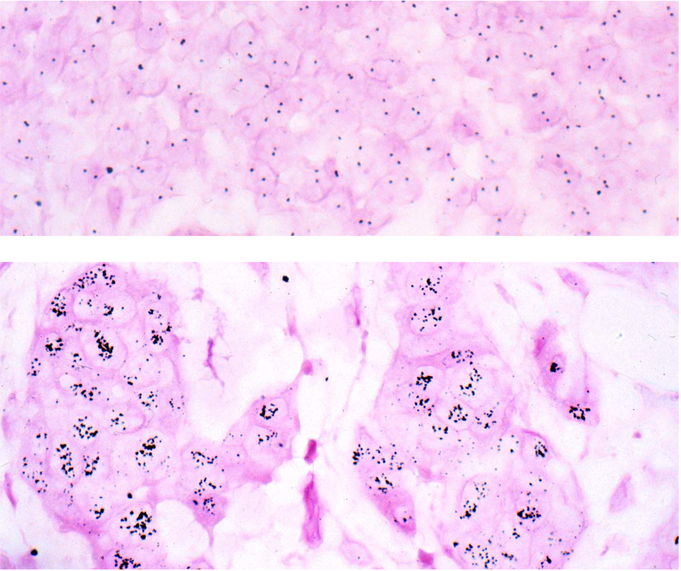
Fig. 14. Her2 breast cancer test. In these human breast biopsies, our sensitive EnzMet detection method was used to visualize the Her2 gene (out of ~25,000 other genes). Top image is from a Her2 negative patient showing the normal 2 genes per cell. The bottom image is from a patient showing the gene duplication that happens in Her2 cancer, with multiple copies in the nuclei.
On Failure...
Our goal of better treating those suffering from cancer has not been achieved. We have tried a number of approaches, but unfortunately unanticipated roadblocks were found.
This slow string of failures has had some benefit in that we have learned more about cancers and pitfalls of various approaches. Along the way we have developed tools that Nanoprobes provides to other researchers, many in the cancer field, and have provided some inspiration to others for further studies.
The Best for Last…
If you have read this far, we are saving, we hope, the best for last. We have been disappointed so many times that the encouraging promise of our most recent attempt to better treat cancers has to be viewed with guarded optimism.
Learning from the problems of high Z metal nanoparticles, poor clearance and discoloration, we have moved to biodegradable protein nanoparticles instead.
Our most recent project has yielded a real breakthrough.
Our most recent project is now entering its fifth year and has yielded a breakthrough way to beat cancer’s malignant behavior by hitchhiking on the body’s own pathways.
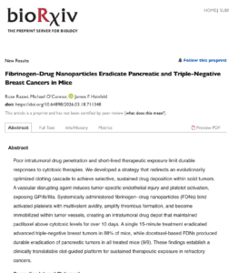
Mice with advanced, otherwise untreatable and deadly cancers have recovered and returned to a normal, cancer-free life. In our current study, we are having amazing results with both triple-negative breast cancer and pancreatic cancer in mice – both notoriously hard to treat. See the preprint of our upcoming paper here
This is the breakthrough we've been waiting for.
Our new multifunctional nanoparticles have these properties:
- After IV injection target tumors wherever they are
- Once in the tumor they polymerize to prevent washout
- This also cuts blood supply killing much of the tumor
- They then release an anti-cancer drug at high levels over about a 2 week period
- They are biodegradable
- They are not immunogenic
- But they make tumors more immunogenic
Our new approach to treating cancer does not depend on the type of cancer. We expect it to work just as well on lung, breast, colon or prostate cancer… in fact, possibly on 90% of all cancers.
With this breakthrough, it really may be possible to cure cancer.
Bringing our breakthrough therapy into the world
See a preprint of our upcoming paper on BioRkiv
Get a sneak peak at our upcoming paper, featuring our latest results.
GIST is a plain-language AI summary service run by a fellow researcher, helping to bring new research to a wider audience.
Is a Human Treatment Available?
Unfortunately, not yet. Cancer, and other human treatments, have to be tested, reviewed, and approved by the FDA. This is to ensure patient safety and benefit. Proving this is expensive and takes time, but is vitally important to protect and ensure benefit to patients. Our goal is to get this to patients safely, effectively, ASAP.
What’s next?
This research produces no income, only expenses. Therefore, financially we need your help to move this breakthrough technology from the laboratory to the bedside to benefit cancer victims, for whom every day is precious.
Project Cure Cancer
To move this exceptionally promising new treatment to human benefit it must be approved by the FDA. The two items for approval are Safety and Efficacy. Before any human trial is allowed, safety must be shown in 2 larger species.
Since Nanoprobes does not have the facilities, this required safety study will be outsourced. The treatment is very well tolerated in mice, so we don’t expect any pain or discomfort to these animals and every attempt is made to use as few animals as possible. Two quotations were approximately the same, $900,000. The treatment solutions must also be prepared in a sterile pharmaceutical level production facility in much greater quantities than those used for mice, and that will cost $500,000. Total cost $1,400,000.
Thank you!