
Precision Nanogold® labels for Click chemistry!
Click chemistry is the reaction between an azide and an alkyne or similar group. It is a bioorthogonal reaction (i.e. it does not occur naturally) which lets let you label selectively in living cells, tissues and even animals without cross-reactivity.
Click 0.8 nm Nanogold® (“Undecagold”) reagents are stable, biocompatible, and well tolerated by living cells. They provide ultra-high resolution, putting the Undecagold right on the target site: locate targets at macromolecular resolution.
Available in the Click chemistry of your choice:
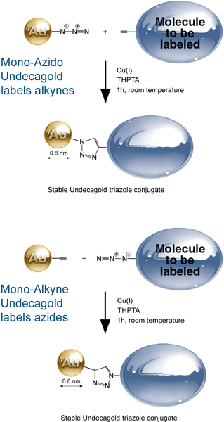
- Mono-Azido Nanogold® reacts with alkynes and related reagents; may be used for some copper-free click reactions.
- Mono-Alkyne Nanogold® reacts with azides.
- Mono-DBCO-Nanogold® has a Dibenzocyclooctyl (DBCO) group for copper-free CLICK labeling with an azide.
Applications:
- Label dynamic processes in living cells or tissues – Click reagents will not hydrolyze or degrade.
- Label components in living cells: do not react with endogenous species like biotin or cross-reactive proteins.
- High-resolution labeling of subunits or molecular sites: just incorporate the corresponding reactive azide or alkyne into the target – no antibody or probe required.
- Optical Visualization: use silver or gold enhancement to visualize by light microscopy or for super-sensitive blotting. See as little as 1 pg of target IgG!
- Multiplex your labeling: Add a click reaction to detect an additional target if there is no suitable antibody or probe.
0.8 nm Nanogold® Labeling Reagents
Ultra-small, 0.8 nm Nanogold® is the best choice for cryo-EM.
“Undecagold” (Au11) is smaller than our original 1.4 nm Nanogold®, with a core of 11 gold atoms only 0.8 nm in diameter. It is ideal for ultra-high-resolution EM work such as cryo-EM, scanning transmission electron microscopy (STEM), or for resolving elements of large structures by TEM in conjunction with image processing.
0.8 nm Nanogold® (“Undecagold”) has been used to see the biotin binding sites on avidin to 1 nm resolution by electron microscopy. It is prepared in a form with one reactive arm for cross-linking to a specific site on a target molecule, and is available with different reactivities for labeling different sites.
Ultra small 0.8 nm gold core
- Use to prepare the smallest possible gold probes.
- Highest possible resolution.
- Site specific covalent labeling with choice of reactivities.
Note: 0.8 nm Nanogold® (Undecagold) is perfect for labeling in high-resolution applications like cryo-EM, but single Undecagold clusters are not routinely visualized directly in the TEM. For standard TEM, Undecagold may be seen upon image processing of protein helices and crystals, or visualized en masse if there is a bulk deposition such as staining of an organelle.
Also, Undecagold develops more slowly and with less final developer deposition during silver enhancement or gold enhancement than with our larger, 1.4 nm Nanogold®. Therefore, for many applications we recommend 1.4 nm Nanogold®.
![Structure of 0.8 nm Nanogold® gold nanoparticle (undecagold) showing the [Au11] core, coordinated ligands and peripheral reactive group (maleimide).](https://nanoprobes.com/wp-content/uploads/catfig9.gif)
Structure of undecagold showing the [Au11] core, coordinated ligands and peripheral reactive group (maleimide).
Easily enhanced for electron microscopy, light microscopy, cryo-EM, blots…
Try our precision nanoparticle developers for easily controlled development with low background!
Applications
- Cryo-EM – use ultra-small Undecagold for your highest-resolution, cryo-electron microscopy applications.
- Scanning transmission electron microscopy (STEM). [References]
- Undecagold labeling identifies fibrinogen cross-linking site. (Mosesson et al., 1998)
- Labeling of F-actin at the Cys-374 residue by reacting with monomalemido undecagold; data was collected from frozen hydrated samples in the EM and image processing used to map the distribution of label
- Milligan, R.A., et al.: Molecular structure of F-actin and location of surface binding sites. Nature, 348, 217-221 (1990)).
- Labeling of a cysteine in cytochrome oxidase crystals and EM visualization in uranyl acetate/glucose using image processing.
- Crum, J., Gruys, K.J., and Frey, T.G. Electron microscopy of cytochrome c oxidase crystals: labeling of subunit III with a monomaleimide undecagold cluster compound. Biochemistry. 1994 Nov 22;33(46):13719-26.
- tRNA labeling with undecagold using a modified nucleic acid base
- Blechschmidt, B., Shirokov, V., and Sprinzl M. Undecagold cluster modified tRNA (Phe) from Escherichia coli and its activity in the protein elongation cycle. J. Biochem., 219, 65-71 (1994). )
- Localization of the C terminus of the assembly domain of hepatitis B virus capsid protein by electron microscopy, with image processing
- Zlotnick, A., Cheng, N., Stahl, S. J., Conway, J. F., Steven, A. C., and Wingfield, P. T.: Localization of the C terminus of the assembly domain of hepatitis B virus capsid protein: Implications for morphogenesis and organization of encapsidated RNA; Proc. Natl. Acad. Sci. USA, 94, 9556-9561 (1997).
- Isomorphous heavy atom replacement in crystallography of large structures by EM crystallography, single particle alignment work, or X-ray diffraction. Provides a signal much greater than a single heavy atom label
- Thygesen, J., Weinstein, S., Franceshi, F., and Yonath, A.: The suitability of multi-metal clusters for phasing in crystallography of large macromolecular assemblies [review]; Structure, 4, 513-518 (1996). [Full PDF available here]
- Labeling carbohydrates on a glycoprotein: sugars are first oxidized to produce aldehydes, which react with the monamino undecagold.
- Lipka, J.J., Hainfeld, J.F., and Wall, J.S. Undecagold labeling of a glycoprotein: STEM visualization of an undecagoldphosphine cluster labeling the carbohydrate sites of human haptoglobin-hemoglobin complex. J. Ultrastruct. Res., 84, 120 (1983).