Fluorescein-FluoroNanogold™ Fab’ goat anti-rabbit
Fluorescent labels PLUS Nanogold® – covalently bound. Make your target visible in almost any microscope!
Fluorescein-FluoroNanogold™ Fab’ goat anti-rabbit
Fluorescent labels PLUS Nanogold® – covalently bound. Make your target visible in almost any microscope!
Fluorescein-FluoroNanogold™ Fab’ goat anti-rabbit
7004-1ML
1.0 mL
$ 630.00
Fluorescein-FluoroNanogold™ Fab’ goat anti-rabbit
7004-0.5ML
0.5 mL
$ 378.00
- 7004-1ML
Fluorescein-FluoroNanogold™ Fab’ goat anti-rabbit
1.0 mL
$ 630.00
- 7004-0.5ML
Fluorescein-FluoroNanogold™ Fab’ goat anti-rabbit
0.5 mL
$ 378.00
Grow the size - Keep the precision
Combine nanoparticle developers with Nanogold® labels
Easy, archival developers for any scope or blots
- GoldEnhance™:
Simple. Superior. Precision nanoparticle enhancement
Sharp, archival staining of blots or easy viewing in any scope. - Silver Enhancement:
Precision nanoparticles, not colloidal silver blobs: precise development for any scope.
Product Information
- Hirano K, Kinoshita T, Uemura T, Motohashi H, Watanabe Y, Ebihara T, Nishiyama H, Sato M, Suga M, Maruyama Y, Tsuji NM, Yamamoto M, Nishihara S, Sato C. Electron microscopy of primary cell cultures in solution and correlative optical microscopy using ASEM. Ultramicroscopy. 2014 Aug;143:52-66. doi: 10.1016/j.ultramic.2013.10.010. Epub 2013 Oct 22. PMID: 24216127.
https://pubmed.ncbi.nlm.nih.gov/24216127/
Fluorescein-FluoroNanogold™ Fab’ goat anti-rabbit
FluoroNanogold™ Combination Labels
Covalently bound, Nanogold® + fluorescent secondary antibodies
Precision meets flexibility
Fluorescein FluoroNanogold™
2-in-1 immunolabeling!
Fluorescein is the most widely used fluorophore; fluorescein FluoroNanogold™conjugates combine fluorescein and Nanogold® in a single convenient, stable probe.
- correlate fluorescein fluorescence staining with brightfield light microscopy and electron microscopy using a single convenient immunostaining procedure.
- Check labeling by fluorescence before EM processing.
- Available in 1 mL or affordable 0.5 mL sizes.
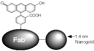
Structure of Fluorescein FluoroNanogold™, showing covalent attachment.
FluoroNanogold™ Staining of Microtubules
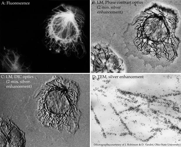
Immunolocalization of microtubules with Fluorescein FluoroNanogold™-Fab’ in the same human monocyte visualized by various microscopies: (A) Fluorescence; (B) Phase; (C) DIC, and (D) Electron Microscopy (with silver enhancement). (Micrographs courtesy of Dr. J. M. Robinson and Dr. D. Vandré, Ohio State University).
Easily enhanced for electron microscopy, light microscopy, cryo-EM, blots…
Try our precision nanoparticle developers for easily controlled development with low background!
FluoroNanogold™ Combination Labels
Covalently bound, Nanogold® + fluorescent secondary antibodies
Precision meets flexibility
Each secondary antibody / Fab’ includes TWO labels
Covalently bound for stability and long working times:
-
Fluorescent dye for imaging in Super-Resolution
-
Nanogold® particles mark your target for TEM, light microscopy and blots!
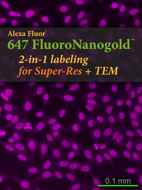
2-in-1 immunolabeling for Super-Res!
Simultaneously label for Super-Res AND TEM / light microscopes in a single, standard immunolabeling procedure
Finally– Put your Super-Res images into cellular context!
- Reveal your fluorescent labeling in traditional microscopes, where you can see the surrounding cell structures!
- TEM, light microscope, phase contrast, etc.
- Adaptable to any resolution, for ultimate flexibility in correlative studies
- Easy, archival enhancement kits grow the Nanogold® as big as you like
!["FNG [FluoroNanogold™] is a probe containing two different markers; it opens the possibility of imaging the same sample at both the optical and the EM level. Thus, it is easy to perform a multimodal investigation, either on different cells or on the same cell visualized by correlative microscopy." Cheutin, T.; Sauvage, C.; Tchélidzé, P, O'Donohue, M. F.; Kaplan, H.; Beorchia, A., and Ploton, D.: Visualizing macromolecules with FluoroNanogold: from photon microscopy to electron tomography. Methods Cell Biol., 79, 559–574 (2007). [This is an entire chapter on FluoroNanogold, providing a detailed and well illustrated write-up of techniques for combined confocal fluorescence microscopic labeling and electron tomography.]](https://nanoprobes.com/wp-content/uploads/Vol11_Iss6_Fig1a-550w.jpg)
“FNG [FluoroNanogold™] is a probe containing two different markers; it opens the possibility of imaging the same sample at both the optical and the EM level. Thus, it is easy to perform a multimodal investigation, either on different cells or on the same cell visualized by correlative microscopy.”
Cheutin, T.; Sauvage, C.; Tchélidzé, P, O’Donohue, M. F.; Kaplan, H.; Beorchia, A., and Ploton, D.: Visualizing macromolecules with FluoroNanogold: from photon microscopy to electron tomography. Methods Cell Biol., 79, 559–574 (2007). [This is an entire chapter on FluoroNanogold, providing a detailed and well illustrated write-up of techniques for combined confocal fluorescence microscopic labeling and electron tomography.]
By combining gold and fluorescence into one immunoprobe, the same specimen may be imaged using both fluorescence microscopy (e.g., with a confocal microscope) and at the ultrastructural level by electron microscopy.
FluoroNanogold™is now available with Alexa Fluor®* 647, 546, 488 or 594, giving you the benefits of brighter fluorescence, reduced photobleaching, and compatibility with a wider pH range, or in its original formulation with fluorescein as the fluorophore.
Unlike colloidal golds which quench fluorescence, tiny Nanogold® lets the fluorescence shine through. All components are covalently attached to ensure stability and long shelf life. Fab’ antibody fragments are much smaller probes than IgG conjugates, and have shown excellent penetration into cells and nuclei. FluoroNanogold™conjugates are chromatographically purified to eliminate any aggregates, free gold or unattached fluorescent molecules.
- Unprecedented correlation between fluorescence and EM data.
- Single labeling procedure means less chance for specimen perturbation.
- Choice of new fluorophores for brighter fluorescence, lower background and multicolor labeling.
- Same excellent penetration as found with Nanogold®: much better than 5 and 10 nm colloidal gold probes.
- FluoroNanogold™ probes are smaller than IgG conjugates (we use Fab’ fragments).
- Covalent coupling of both labels gives stability and long shelf life.
- Conjugates are stable and fluorescent at a wide range of pH and ionic strengths.
Applications
- Correlative fluorescence and electron microscopy
- Check your labeling by fluorescence before processing for EM
- Monitor a dynamic process by fluorescence to determine when to fix and process for EM.
- Differentiate different targets using different fluorophores.
Immunolocalization of microtubules with Fluorescein FluoroNanogold™-Fab’ in the same human monocyte visualized by various microscopies: (A) Fluorescence; (B) Phase; (C) DIC, and (D) Electron Microscopy (with silver enhancement). (Micrographs courtesy of Dr. J. M. Robinson and Dr. D. Vandré, Ohio State University).
See your target’s fluorescent label as before
PLUS the Nanogold label for Super-Res, LM, EM and blots
Featured paper:
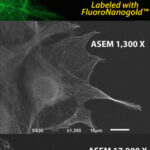
Sato et al. (2013) used FluoroNanogold™ to explore the mechanism behind cellular dynamics, using correlative microscopy with fluorescence and the new Atmospheric SEM (ASEM).
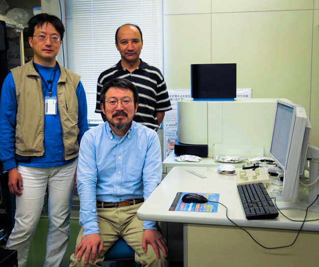
Dr. Chikara Sato and colleagues with the Atmospheric Scanning Electron Microscope (ASEM).
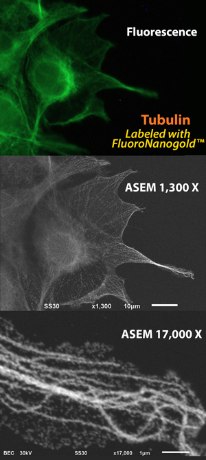
Fig. 1 Microtubules, visualized by optical microscopy (OM) and atmospheric scanning electron microscopy (ASEM: inverted SEM). COS7 cells were labeled with anti-alpha-tubulin primary antibody followed by Alexa Fluor 488 FluoroNanogold™ secondary antibody (Nanoprobes). Top: Fluorescence microscopy image. Middle: ASEM image of the same cells after gold enhancement by GoldEnhance™ EM (Nanoprobes). Botton: Higher magnification image of the right filopodia. Microtubule rails are clearly observed in the cytoplasm. (Sato et al., 2013)