Updated: Vebruary 3, 2003
N A N O P R O B E S E - N E W S
Vol. 4, No. 2 February 3, 2003
This monthly newsletter is to keep you informed about techniques to improve your immunogold labeling, highlight interesting articles and novel metal nanoparticle applications, and answer your questions. We hope you enjoy it and find it useful.
Have questions, or issues you would like to see addressed in the next issue? Let us know by e-mailing tech@nanoprobes.com.
. . . . . . . . . . . . . . . . . . . . . . . . . . . . . . . .
Nanogold is thought to be mostly a discrete chemical compound, rather than a colloidal particle. Therefore, its extinction coefficients are well-defined, and these may be used to determine the extent of labeling - so you can calculate how many Nanogold particles have bound per antibody, protein, or other conjugate biomolecule.
Gold labeling of most proteins is best calculated using the extinction coefficients at 280 nm, where proteins usually absorb strongly and their extinction coefficients are known, and 420 nm, where Nanogold® absorbs strongly but most proteins do not. If your protein does not absorb at 420 nm, use the following method to calculate labeling:
- Since the absorption at 420 nm arises solely from the Nanogold, use the extinction coefficient of Nanogold at 420 nm (110,000) to calculate the concentration of Nanogold:
Nanogold concentration = A420nm / 110,000 mol/l
- Use the absorption at 420 nm and the extinction coefficients of Nanogold at 280 nm and 420 nm to calculate the absorption at 280 nm due to the Nanogold:
A280nm of Nanogold = A420nm x 300,000 / 110,000
- Subtract this from the measured absorption at 280 nm. The difference is the absorption due to protein:
A(protein) = A(measured) - A(Nanogold)
- Use the absorption arising from protein to calculate the protein concentration:
Protein concentration = A(protein) / E(protein)
where E is the extinction coefficient of the protein at 280 nm. You can convert optical densities to extinction coefficients using the molecular weight as follows:
E = OD (1%) X MW(protein) / 10
- The number of Nanogold labels per protein is simply the concentration of Nanogold divided by the concentration of protein:
Labeling = Nanogold concentration / protein concentration
The spectroscopic basis for this calculation, as well as the spectra of Nanogold and undecagold, are given in our Guide to Gold Cluster Labeling. If you are labeling a molecule that is quantitated by its absorption at a different wavelength - for example DNA, which is quantitated using absorption at 260 nm - you can use these spectra to estimate the extinction coefficient of Nanogold or undecagold at this wavelength and so calculate labeling.
Some extinction coefficients commonly used for labeling calculations:
| Molecule |
280 nm |
420 nm |
| Nanogold |
300,000 |
110,000 |
| Undecagold |
168,000 |
47,100 |
| IgG |
225,000 |
~ 0 |
| Fab' |
75,000 |
~ 0 |
Further Information:
If your biomolecule also absorbs at 420 nm, you will need to use a slightly longer matrix-based method to calculate labeling. This is given in full on a separate page of the Guide:
. . . . . . . . . . . . . . . . . . . . . . . . . . . . . . . .
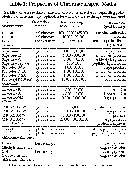
Before you can accurately determine labeling, you need to separate the conjugate from the other species in the reaction mixture, particularly excess unconjugated Nanogold or unlabeled biomolecule. You can ensure an efficient separation not just be picking the right separation method, but also by choosing a ratio of gold label to biomolecule such that of the two reagents, the one that is more easily separated if it is unreacted is in excess, and the less easily separated one is the limiting reagent and is completely conjugated.
We usually recommend liquid chromatography over a gel filtration or size fractionation column for conjugate separation. In this system, the more easily separated component will be the smaller of the two, and therefore this should be present in excess. If you are labeling a large protein such as an IgG molecule, the gold label should be present in excess; if the size difference is great, use a larger excess (3-fold or greater), but if it is less (for example, a large peptide with a MW of 25,000) use a smaller excess (2-fold). If the biomolecule you are labeling is smaller that the gold label, use an excess of the biomolecule; again, use a larger excess (10-fold or greater) if the size difference is great, but a smaller excess (2-fold to 5-fold) if the biomolecule is only slightly smaller than the gold label.
Approximate molecular weights of gold cluster labels:
- Nanogold: 15,000
- Undecagold: 5,000
Note, however, that because a significant contribution to MW is made by the very dense gold core, these molecules are smaller than proteins of equivalent molecular weight. We find that Nanogold behaves similarly to a protein of MW 8 - 10,000.
More information:
Other chromatographic methods may be helpful. If your conjugate biomolecule is charged or has very distinct hydrophobic properties, then ion exchange, hydrophobic interaction, or reverse-phase chromatography may also be helpful.
To reduce your reaction volumes for injection onto columns, we recommend using a centrifuge membrane filter, such as the Centricon (available from Millipore); choosing the right MW cutoff can help separation - 30,000 cut-off works well for larger proteins, and 10,000 is good for smaller molecules.
. . . . . . . . . . . . . . . . . . . . . . . . . . . . . . . .
(1) Gel electrophoreis:
We are frequently asked whether Nanogold®-labeled molecules may be separated using gel electrophoresis. While there is evidence that this may be useful, we advise caution in interpreting the results, as gel shifts alone are frequently not found to be good indicators of labeling. Although Nanogold has a nominal MW of 15,000, other properties can contribute to the mobility of compounds, and in practice the observed gel shift may be very small.
If you wish to use this method, we recommend the following:
- Use non-reducing gels: thiol-containing reagents such as dithiothreitol (DTT) can break down the Nanogold cluster.
- Detect each component separately and specifically to confirm labeling. Separate the reaction mixture or product, divide the gel, then stain one portion with silver enhancement (note: do not use silver protein stain, but a silver enhancement reagent specific for gold particles, such as LI Silver from Nanoprobes) or gold enhancement, and the other with a general protein stain such as Coomassie blue. You may be able to resolve two bands, one which develops with silver enhancement *and* Coomassie (the Nanogold conjugate) and one of which develops only with Coomassie (unlabeled protein). The presence of unbound Nanogold would add a third band, which reacts only with silver.
- When silver or gold enhancing to detect the Nanogold conjugate, monitor the progress of development carefully. This is a highly sensitive detection method, and in addition to the target band, a "tail" may also develop behind it, which, if left too long, may obstruct resolution of the band.
For some illustrations, see p. 92-94 of Hainfeld and Furuya:
Hainfeld, J. F., and Furuya, F. R.: Silver Enhancement of Nanogold and Undecagold. In "Immunogold-Silver Staining: Principles, Methods and Applications," M. A. Hayat (Ed.); CRC Press, Boca Raton, FL, 1995, pp. 71-96.
An application note, "Detection of Nanogold-labeled molecules on Gels," is available on our web site. More information:
(2) Dialysis:
Dialysis offers an attractively simple method in theory. However, results obtained in practice do not always match those predicted, and large losses of both gold label and conjugate biomolecule have occasionally been observed. If you wish to try dialysis, we recommend the following:
- Select a tubing material that does not contain any materials that might either bind to Nanogold or break down the gold cluster core. Such materials include thiol-containing compounds, and also hydrophobic materials, which may adhere to the cluster surface.
- Test the membrane with a small amount of unconjugated Nanogold before attempting to separate your conjugate. This will indicate both whether the membrane is compatible with the gold label, and whether its permeability properties match those desired.
Density gradient ultracentifugation is often used for separation of colloidal gold conjugates. However, we have found that generally, Nanogold and undecagold are too small to be easily separated by this method.
. . . . . . . . . . . . . . . . . . . . . . . . . . . . . . . .
Nanoprobes offers two negative stain reagents with complementary properties. NanoVan is recommended for use with Nanogold® because it is based on vanadium and is therefore lighter than heavy metal based stains such as uranyl acetate or lead citrate, and hence makes visualization easier. Nano-W is based on the heavier element tungsten and therefore gives a more dense stain. These two reagents are completely miscible, and therefore may be mixed together in different proportions to control the density of staining more precisely.
Both have near-neutral pH, and NanoVan has been found to be less susceptible to electron beam damage than uranyl acetate. For example, Franzetti and colleagues used negative stain electron microscopy with NanoVan, and image analysis of 1,300 particles, to define the structure of a large, tetrahedral, dodecameric protease complex (TET) from archaea. TET, which has broad aminopeptidase activity and can process peptides of up to 30-35 amino acids in length, has a central cavity accessible through four narrow channels (<17 Å wide) and four wider channels (21 Å wide). The authors found NanoVan's pH of close to 8 was better suited to their system than that of uranyl acetate.
Reference:
Franzetti, B.; Schoehn, G.; Hernandez, J. F.; Jaquinod, M.; Ruigrok, R. W.; and Zaccai, G.: Tetrahedral aminopeptidase: a novel large protease complex from archaea. EMBO J., 21, 2132-2138 (2002).
Abstract (courtesy of the EMBO Journal):
http://emboj.oupjournals.org/cgi/content/abstract/21/9/2132
Fry and co-workers used Nano-W with 6 nm protein-A gold in the molecular characterization of a novel human centrosomal protein, C-Nap1 (for centrosomal Nek2-associated protein 1); immunoelectron microscopy revealed that C-Nap1 is associated specifically with the proximal ends of both mother and daughter centrioles. Whereas C-Nap1 was concentrated at centrosomes in all interphase cells, immunoreactivity at mitotic spindle poles was strongly diminished, and based on these and other data, a model implicating both Nek2 and C-Nap1 in the regulation of centriolecentriole cohesion during the cell cycle was proposed.
Negative staining immunoelectron microscopy of isolated centrosomes was performed by diluting centrosomes 1:10 in PBS before sedimentation onto Pioloform and carbon-coated grids using an airfuge (65,000 g-av, 15 min). Grids were blocked with 0.1% gelatin in PBS, incubated with affinity-purified antiC-Nap1 IgGs (R63, 1 microgram/ml in blocking buffer, 45 min) and protein A6-nm gold, then negatively stained with Nano-W.
Reference:
Fry, A. M.; Mayor, T.; Meraldi, P.; Stierhof, Y.-D.; Tanaka, K., and Nigg, E. A.: C-Nap1, a novel centrosomal coiled-coil protein and candidate substrate of the cell cycle-regulated protein kinase Nek2. J. Cell Biol., 141, 1563-74 (1998).
Abstract (courtesy of the Journal of Cell Biology):
http://www.jcb.org/cgi/content/abstract/141/7/1563
Reprint (courtesy of the Journal of Cell Biology):
http://www.jcb.org/cgi/reprint/141/7/1563.pdf
More information:
. . . . . . . . . . . . . . . . . . . . . . . . . . . . . . . .
Liu and co-workers recently used Nanogold® to label the outside of DNA nanotubes; this was accomplished by incorporating an amino- group into the nanotube-forming oligonucleotides such that it was positioned on the surface of the resulting nanotube, then reacting with Mono-Sulfo-NHS-Nanogold. However, nanotubes assembled from 3' and 5'-thiolated oligonucleotides did not react with Monomaleimido-Nanogold, indicating that the thiols were buried within the nanotubes.
Reference:
Liu, D.; Reif, J., and LaBean, T: DNA Nanotubes: Construction and Characterization of Filaments Composed of TX-tile Lattice. Lecture Notes in Computer Science #2568: 8th International Workshop on DNA-Based Computers, DNA8 Sapporo, Japan, June 10-13, 2002, p. 10ff (2003).
Abstract (courtesy of Springer):
http://link.springer.de/link/service/series/0558/bibs/2568/25680010.htm
Keren and group have demonstrated specific localization of biotinylated DNA probes on DNA substrates with different junction topologies using Nanogold-streptavidin, and observed the Nanogold by atomic force microscopy (AFM) both before and during autometallography, thus demonstrating an approach to sequence-specific molecular lithography.
Reference:
Keren, K.; Krueger, M.; Gilad, R.; Ben-Yoseph, G.; Sivan, U., and Braun, E.: Sequence-specific molecular lithography on single DNA molecules. Science, 297, 72-75 (2002).
Abstract (Medline):
http://www.ncbi.nlm.nih.gov:80/entrez/query.fcgi?cmd=Retrieve&db=PubMed&list_uids=12098693&dopt=Abstract
Yano and co-workers have developed an antigen retrieval procedure that enables the immunoelecton microscopic labeling of chromogranin in secretory granules, thus allowing the identification of secretory granules. Ultrathin epoxy-embedded sections that were microwaved in alkaline solution (pH 10) were adequately labeled with 15 nm gold-conjugated secondary antibodies (5.2 ± 0.34 particles per SG). Considerably improved efficiency of immunostaining was achieved by microwaving sections in alkaline solution (pH 10) with subsequent immunostaining at 60°C (12.2 ± 0.51 particles per SG); the method was also applied to epoxy-embedded sections obtained from formalin-fixed, paraffin-embedded blocks of tissue and was even valid for an old epoxy-embedded block prepared 15 years previously.
Reference:
Yano, S.; Kashima, K.; Daa, T.; Urabe, S.; Tsuji, K.; Nakayama, I., and Yokoyama S.: An Antigen Retrieval Method Using an Alkaline Solution Allows Immunoelectron Microscopic Identification of Secretory Granules in Conventional Epoxy-embedded Tissue Sections. J. Histochem.Cytochem., 51, 199-204 (2002).
Abstract (courtesy of the Journal of Histochemistry and Cytochemistry):
http://www.jhc.org/cgi/content/abstract/51/2/199
. . . . . . . . . . . . . . . . . . . . . . . . . . . . . . . .
To unsubscribe from this newsletter, simply reply to this message, and replace the subject line with "unsubscribe." Alternatively, visit our web site newsletter page at www.nanoprobes.com/Newsletter.html#unsubscribe.
|